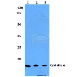
Figure 1: Western Blot - Anti-Cystatin A Antibody [ARC1993] (A309340)
Western blot analysis of extracts of various cell lines, using Anti-Cystatin A Antibody [ARC1993] (A309340) at 1:1,000 dilution. The secondary antibody was Goat Anti-Rabbit IgG H&L Antibody (HRP) at 1:10,000 dilution. Lysates/proteins were present at 25µg per lane. The blocking buffer used was 3% non-fat dry milk in TBST. Detection was with a ECL Enhanced Kit (RM00021). Exposure time: 180s.
Figure 2: Immunohistochemistry - Anti-Cystatin A Antibody [ARC1993] (A309340)
Immunohistochemistry analysis of paraffin-embedded rat lung using Anti-Cystatin A Antibody [ARC1993] (A309340) at a dilution of 1:100 (40x lens). Perform microwave antigen retrieval with 10 mM Tris/EDTA buffer pH 9.0 before commencing with IHC staining protocol.
Figure 3: Immunohistochemistry - Anti-Cystatin A Antibody [ARC1993] (A309340)
Immunohistochemistry analysis of paraffin-embedded human colon carcinoma tissue using Anti-Cystatin A Antibody [ARC1993] (A309340) at a dilution of 1:100 (40x lens). Perform microwave antigen retrieval with 10 mM Tris/EDTA buffer pH 9.0 before commencing with IHC staining protocol.
Figure 4: Immunofluorescence - Anti-Cystatin A Antibody [ARC1993] (A309340)
Immunofluorescence analysis of NIH-3T3 cells using Anti-Cystatin A Antibody [ARC1993] (A309340) at a dilution of 1:100 (40x lens). DAPI was used to stain the cell nuclei (blue).
Publishing research using Anti-Cystatin A Antibody [ARC1993] (A309340)? Please let us know so that we can list the citation on this page.
Alternative products to Anti-Cystatin A Antibody [ARC1993] (A309340)


![Western Blot - Anti-Cystatin A Antibody [ARC1993] (A309340) - Antibodies.com](https://cdn.antibodies.com/image/catalog/309/A309340_1.jpg?profile=product_top)
![Immunohistochemistry - Anti-Cystatin A Antibody [ARC1993] (A309340) - Antibodies.com](https://cdn.antibodies.com/image/catalog/309/A309340_2.jpg?profile=product_top)
![Immunohistochemistry - Anti-Cystatin A Antibody [ARC1993] (A309340) - Antibodies.com](https://cdn.antibodies.com/image/catalog/309/A309340_3.jpg?profile=product_top)
![Immunofluorescence - Anti-Cystatin A Antibody [ARC1993] (A309340) - Antibodies.com](https://cdn.antibodies.com/image/catalog/309/A309340_4.jpg?profile=product_top)
![Western Blot - Anti-Cystatin A Antibody [ARC1993] (A309340) - Antibodies.com](https://cdn.antibodies.com/image/catalog/309/A309340_1.jpg?profile=product_top_thumb)
![Immunohistochemistry - Anti-Cystatin A Antibody [ARC1993] (A309340) - Antibodies.com](https://cdn.antibodies.com/image/catalog/309/A309340_2.jpg?profile=product_top_thumb)
![Immunohistochemistry - Anti-Cystatin A Antibody [ARC1993] (A309340) - Antibodies.com](https://cdn.antibodies.com/image/catalog/309/A309340_3.jpg?profile=product_top_thumb)
![Immunofluorescence - Anti-Cystatin A Antibody [ARC1993] (A309340) - Antibodies.com](https://cdn.antibodies.com/image/catalog/309/A309340_4.jpg?profile=product_top_thumb)
![Western Blot - Anti-Cystatin A Antibody [ARC1993] (A309340) - Antibodies.com](https://cdn.antibodies.com/image/catalog/309/A309340_1.jpg?profile=product_image)
![Immunohistochemistry - Anti-Cystatin A Antibody [ARC1993] (A309340) - Antibodies.com](https://cdn.antibodies.com/image/catalog/309/A309340_2.jpg?profile=product_image)
![Immunohistochemistry - Anti-Cystatin A Antibody [ARC1993] (A309340) - Antibodies.com](https://cdn.antibodies.com/image/catalog/309/A309340_3.jpg?profile=product_image)
![Immunofluorescence - Anti-Cystatin A Antibody [ARC1993] (A309340) - Antibodies.com](https://cdn.antibodies.com/image/catalog/309/A309340_4.jpg?profile=product_image)
![Immunohistochemistry - Anti-Cystatin A Antibody [CPTC-CSTA-1] (A248281) - Antibodies.com](https://cdn.antibodies.com/image/catalog/248/A248281_1.jpg?profile=product_alternative)
![Immunohistochemistry - Anti-Cystatin A Antibody [CPTC-CSTA-1] - BSA and Azide free (A251463) - Antibodies.com](https://cdn.antibodies.com/image/catalog/251/A251463_1.jpg?profile=product_alternative)
![Immunohistochemistry - Anti-Cystatin A Antibody [CSTA/3553] (A248283) - Antibodies.com](https://cdn.antibodies.com/image/catalog/248/A248283_1.jpg?profile=product_alternative)
![Immunohistochemistry - Anti-Cystatin A Antibody [CSTA/3553] - BSA and Azide free (A251465) - Antibodies.com](https://cdn.antibodies.com/image/catalog/251/A251465_1.jpg?profile=product_alternative)
![Immunohistochemistry - Anti-Cystatin A Antibody [CSTA/2882] (A248282) - Antibodies.com](https://cdn.antibodies.com/image/catalog/248/A248282_1.jpg?profile=product_alternative)
![Immunohistochemistry - Anti-Cystatin A Antibody [CSTA/2882] - BSA and Azide free (A251464) - Antibodies.com](https://cdn.antibodies.com/image/catalog/251/A251464_1.jpg?profile=product_alternative)