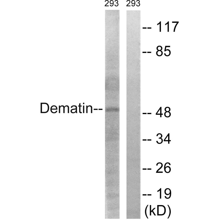
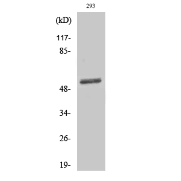
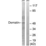
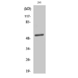
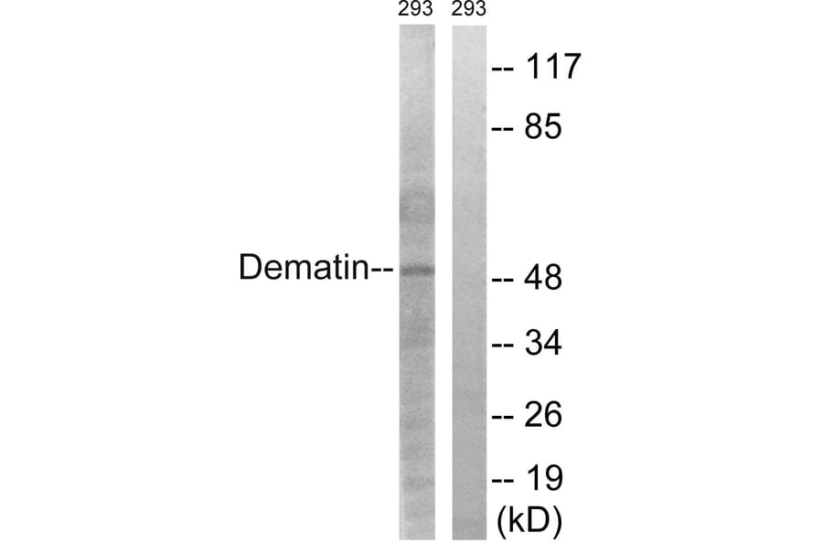
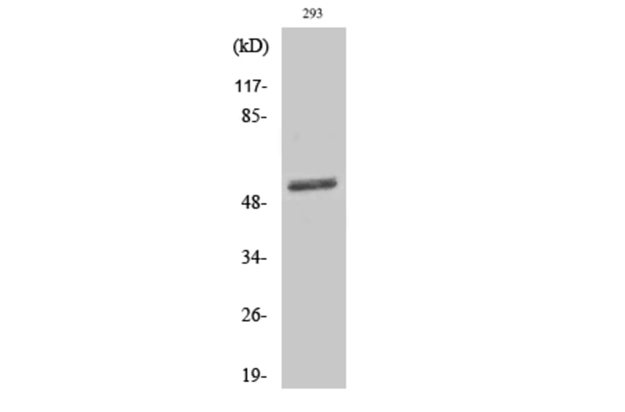
Western blot analysis of lysates from 293 cells using Anti-Dematin Antibody. The right hand lane represents a negative control, where the antibody is blocked by the immunising peptide.
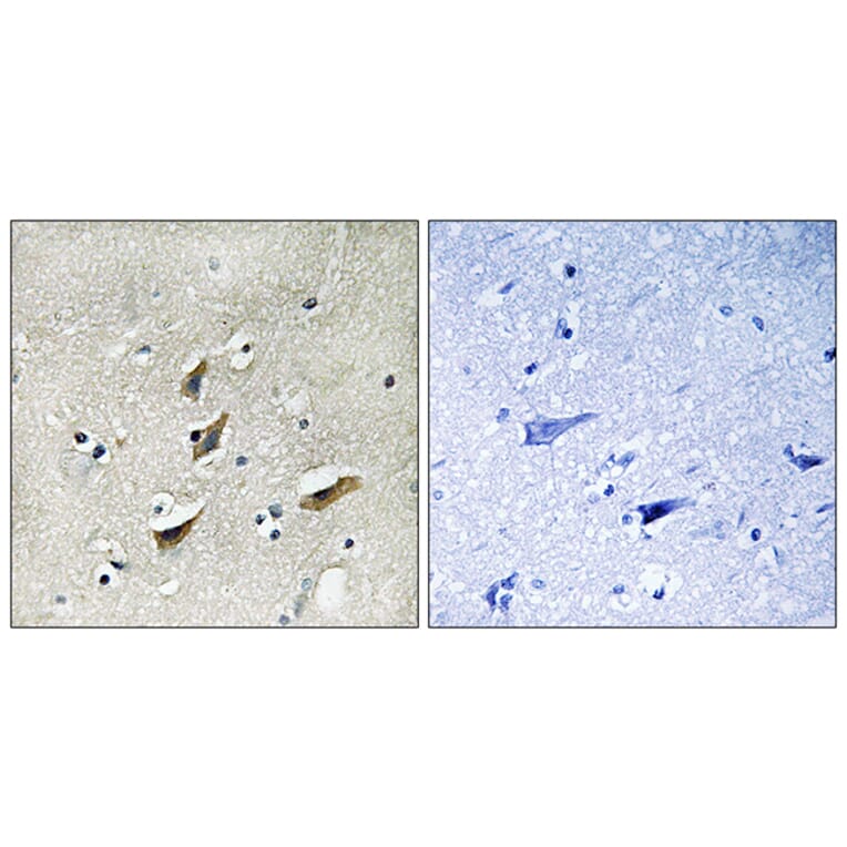
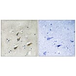
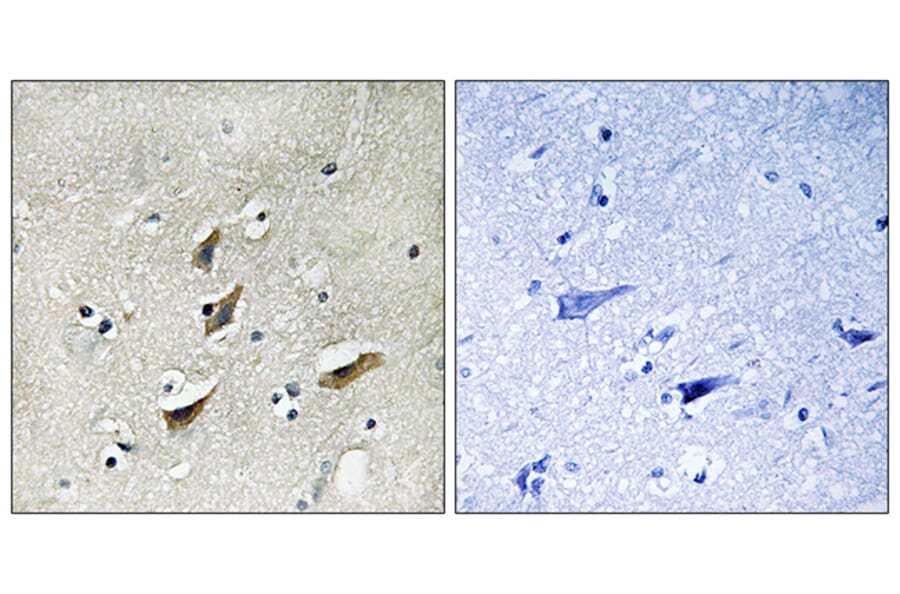
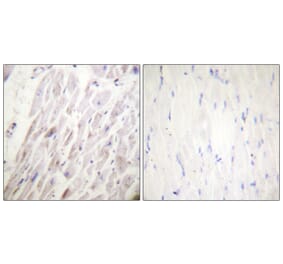
Immunohistochemical analysis of paraffin-embedded human brain tissue using Anti-Dematin Antibody. The right hand panel represents a negative control, where the antibody was pre-incubated with the immunising peptide.
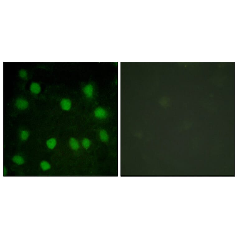
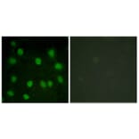
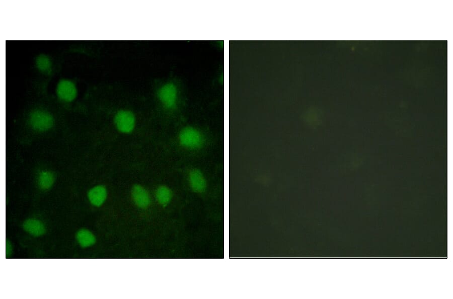
Immunofluorescence analysis of HUVEC cells using Anti-Dematin Antibody. The right hand panel represents a negative control, where the antibody was pre-incubated with the immunising peptide.