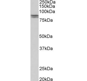
Figure 1: Western Blot - Anti-E Cadherin Antibody [ARC51007 + ARC57093] (A309713)
Western blot analysis of Mouse lung, using Anti-E Cadherin Antibody [ARC51007 + ARC57093] (A309713) at 1:1,000 dilution. The secondary antibody was Goat Anti-Rabbit IgG H&L Antibody (HRP) at 1:10,000 dilution. Lysates/proteins were present at 25µg per lane. The blocking buffer used was 3% non-fat dry milk in TBST. Detection was with a ECL Basic Kit. Exposure time: 30s.
Figure 2: Western Blot - Anti-E Cadherin Antibody [ARC51007 + ARC57093] (A309713)
Western blot analysis of T-47D, using Anti-E Cadherin Antibody [ARC51007 + ARC57093] (A309713) at 1:1,000 dilution. The secondary antibody was Goat Anti-Rabbit IgG H&L Antibody (HRP) at 1:10,000 dilution. Lysates/proteins were present at 25µg per lane. The blocking buffer used was 3% non-fat dry milk in TBST. Detection was with a ECL Basic Kit. Exposure time: 1s.
Immunohistochemistry analysis of paraffin-embedded human colon carcinoma tissue using Anti-E Cadherin Antibody [ARC51007 + ARC57093] (A309713) at a dilution of 1:200 (40x lens). Perform high pressure antigen retrieval with 10 mM citrate buffer pH 6.0 before commencing with IHC staining protocol.
Immunohistochemistry analysis of paraffin-embedded human kidney using Anti-E Cadherin Antibody [ARC51007 + ARC57093] (A309713) at a dilution of 1:200 (40x lens). Perform high pressure antigen retrieval with 10 mM citrate buffer pH 6.0 before commencing with IHC staining protocol.
Immunofluorescence analysis of MCF7 using Anti-E Cadherin Antibody [ARC51007 + ARC57093] (A309713) at a dilution of 1:400 (40x lens). DAPI was used to stain the cell nuclei (blue).


![Western Blot - Anti-E Cadherin Antibody [ARC51007 + ARC57093] (A309713) - Antibodies.com](https://cdn.antibodies.com/image/catalog/309/A309713_1.jpg?profile=product_top)
![Western Blot - Anti-E Cadherin Antibody [ARC51007 + ARC57093] (A309713) - Antibodies.com](https://cdn.antibodies.com/image/catalog/309/A309713_2.jpg?profile=product_top)
![Immunohistochemistry - Anti-E Cadherin Antibody [ARC51007 + ARC57093] (A309713) - Antibodies.com](https://cdn.antibodies.com/image/catalog/309/A309713_3.jpg?profile=product_top)
![Immunohistochemistry - Anti-E Cadherin Antibody [ARC51007 + ARC57093] (A309713) - Antibodies.com](https://cdn.antibodies.com/image/catalog/309/A309713_4.jpg?profile=product_top)
![Immunofluorescence - Anti-E Cadherin Antibody [ARC51007 + ARC57093] (A309713) - Antibodies.com](https://cdn.antibodies.com/image/catalog/309/A309713_5.jpg?profile=product_top)
![Western Blot - Anti-E Cadherin Antibody [ARC51007 + ARC57093] (A309713) - Antibodies.com](https://cdn.antibodies.com/image/catalog/309/A309713_1.jpg?profile=product_top_thumb)
![Western Blot - Anti-E Cadherin Antibody [ARC51007 + ARC57093] (A309713) - Antibodies.com](https://cdn.antibodies.com/image/catalog/309/A309713_2.jpg?profile=product_top_thumb)
![Immunohistochemistry - Anti-E Cadherin Antibody [ARC51007 + ARC57093] (A309713) - Antibodies.com](https://cdn.antibodies.com/image/catalog/309/A309713_3.jpg?profile=product_top_thumb)
![Immunohistochemistry - Anti-E Cadherin Antibody [ARC51007 + ARC57093] (A309713) - Antibodies.com](https://cdn.antibodies.com/image/catalog/309/A309713_4.jpg?profile=product_top_thumb)
![Immunofluorescence - Anti-E Cadherin Antibody [ARC51007 + ARC57093] (A309713) - Antibodies.com](https://cdn.antibodies.com/image/catalog/309/A309713_5.jpg?profile=product_top_thumb)

![Western Blot - Anti-E Cadherin Antibody [ARC51007 + ARC57093] (A309713) - Antibodies.com](https://cdn.antibodies.com/image/catalog/309/A309713_1.jpg?profile=product_image)
![Western Blot - Anti-E Cadherin Antibody [ARC51007 + ARC57093] (A309713) - Antibodies.com](https://cdn.antibodies.com/image/catalog/309/A309713_2.jpg?profile=product_image)
![Immunohistochemistry - Anti-E Cadherin Antibody [ARC51007 + ARC57093] (A309713) - Antibodies.com](https://cdn.antibodies.com/image/catalog/309/A309713_3.jpg?profile=product_image)
![Immunohistochemistry - Anti-E Cadherin Antibody [ARC51007 + ARC57093] (A309713) - Antibodies.com](https://cdn.antibodies.com/image/catalog/309/A309713_4.jpg?profile=product_image)
![Immunofluorescence - Anti-E Cadherin Antibody [ARC51007 + ARC57093] (A309713) - Antibodies.com](https://cdn.antibodies.com/image/catalog/309/A309713_5.jpg?profile=product_image)
![Immunohistochemistry - Anti-E Cadherin Antibody [CDH1/1525] (A250825) - Antibodies.com](https://cdn.antibodies.com/image/catalog/250/A250825_1.jpg?profile=product_alternative)
![Immunohistochemistry - Anti-E Cadherin Antibody [CDH1/1525] - BSA and Azide free (A254005) - Antibodies.com](https://cdn.antibodies.com/image/catalog/254/A254005_1.jpg?profile=product_alternative)
![Immunohistochemistry - Anti-E Cadherin Antibody [CDH1/2208R] (A250830) - Antibodies.com](https://cdn.antibodies.com/image/catalog/250/A250830_1.jpg?profile=product_alternative)
![Immunohistochemistry - Anti-E Cadherin Antibody [CDH1/2208R] - BSA and Azide free (A254010) - Antibodies.com](https://cdn.antibodies.com/image/catalog/254/A254010_1.jpg?profile=product_alternative)
![Immunohistochemistry - Anti-E Cadherin Antibody [4A2] (A250824) - Antibodies.com](https://cdn.antibodies.com/image/catalog/250/A250824_1.jpg?profile=product_alternative)
![Immunohistochemistry - Anti-E Cadherin Antibody [4A2] - BSA and Azide free (A254004) - Antibodies.com](https://cdn.antibodies.com/image/catalog/254/A254004_1.jpg?profile=product_alternative)
![Immunohistochemistry - Anti-E Cadherin Antibody [CDH1/4585] (A250823) - Antibodies.com](https://cdn.antibodies.com/image/catalog/250/A250823_1.jpg?profile=product_alternative)
![Immunohistochemistry - Anti-E Cadherin Antibody [CDH1/4585] - BSA and Azide free (A254003) - Antibodies.com](https://cdn.antibodies.com/image/catalog/254/A254003_1.jpg?profile=product_alternative)
![SDS-PAGE - Anti-E Cadherin Antibody [Research Grade Biosimilar] - Low endotoxin, Azide free (A324002) - Antibodies.com](https://cdn.antibodies.com/image/catalog/324/A324002_1.jpg?profile=product_alternative)
![Immunohistochemistry - Anti-E Cadherin Antibody [rCDH1/1525] (A250828) - Antibodies.com](https://cdn.antibodies.com/image/catalog/250/A250828_1.jpg?profile=product_alternative)
![Immunohistochemistry - Anti-E Cadherin Antibody [rCDH1/1525] - BSA and Azide free (A254008) - Antibodies.com](https://cdn.antibodies.com/image/catalog/254/A254008_1.jpg?profile=product_alternative)
![Immunohistochemistry - Anti-E Cadherin Antibody [SPM381] - BSA and Azide free (A254006) - Antibodies.com](https://cdn.antibodies.com/image/catalog/254/A254006_1.jpg?profile=product_alternative)