Synonyms
Arc 1, CADH1_HUMAN, Cadherin 1, cadherin 1 type 1 E-cadherin, Cadherin1, CAM 120/80, CD 234, CD 324, CD324, CD324 antigen, CDH1, CDHE, E-Cad/CTF3, E-cadherin, ECAD , Epithelial cadherin, epithelial calcium dependant adhesion protein, LCAM, Liver cell adhesion molecule, UVO, Uvomorulin


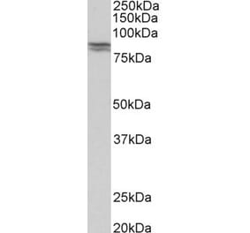
![Western Blot - Anti-E Cadherin Antibody [ARC51012] (A308267) - Antibodies.com](https://cdn.antibodies.com/image/catalog/308/A308267_1.jpg?profile=product_top)
![Western Blot - Anti-E Cadherin Antibody [ARC51012] (A308267) - Antibodies.com](https://cdn.antibodies.com/image/catalog/308/A308267_2.jpg?profile=product_top)
![Immunohistochemistry - Anti-E Cadherin Antibody [ARC51012] (A308267) - Antibodies.com](https://cdn.antibodies.com/image/catalog/308/A308267_3.jpg?profile=product_top)
![Immunohistochemistry - Anti-E Cadherin Antibody [ARC51012] (A308267) - Antibodies.com](https://cdn.antibodies.com/image/catalog/308/A308267_4.jpg?profile=product_top)
![Immunofluorescence - Anti-E Cadherin Antibody [ARC51012] (A308267) - Antibodies.com](https://cdn.antibodies.com/image/catalog/308/A308267_5.jpg?profile=product_top)
![Western Blot - Anti-E Cadherin Antibody [ARC51012] (A308267) - Antibodies.com](https://cdn.antibodies.com/image/catalog/308/A308267_1.jpg?profile=product_top_thumb)
![Western Blot - Anti-E Cadherin Antibody [ARC51012] (A308267) - Antibodies.com](https://cdn.antibodies.com/image/catalog/308/A308267_2.jpg?profile=product_top_thumb)
![Immunohistochemistry - Anti-E Cadherin Antibody [ARC51012] (A308267) - Antibodies.com](https://cdn.antibodies.com/image/catalog/308/A308267_3.jpg?profile=product_top_thumb)
![Immunohistochemistry - Anti-E Cadherin Antibody [ARC51012] (A308267) - Antibodies.com](https://cdn.antibodies.com/image/catalog/308/A308267_4.jpg?profile=product_top_thumb)
![Immunofluorescence - Anti-E Cadherin Antibody [ARC51012] (A308267) - Antibodies.com](https://cdn.antibodies.com/image/catalog/308/A308267_5.jpg?profile=product_top_thumb)

![Western Blot - Anti-E Cadherin Antibody [ARC51012] (A308267) - Antibodies.com](https://cdn.antibodies.com/image/catalog/308/A308267_1.jpg?profile=product_image)
![Western Blot - Anti-E Cadherin Antibody [ARC51012] (A308267) - Antibodies.com](https://cdn.antibodies.com/image/catalog/308/A308267_2.jpg?profile=product_image)
![Immunohistochemistry - Anti-E Cadherin Antibody [ARC51012] (A308267) - Antibodies.com](https://cdn.antibodies.com/image/catalog/308/A308267_3.jpg?profile=product_image)
![Immunohistochemistry - Anti-E Cadherin Antibody [ARC51012] (A308267) - Antibodies.com](https://cdn.antibodies.com/image/catalog/308/A308267_4.jpg?profile=product_image)
![Immunofluorescence - Anti-E Cadherin Antibody [ARC51012] (A308267) - Antibodies.com](https://cdn.antibodies.com/image/catalog/308/A308267_5.jpg?profile=product_image)
![Immunohistochemistry - Anti-E Cadherin Antibody [CDH1/1525] (A250825) - Antibodies.com](https://cdn.antibodies.com/image/catalog/250/A250825_1.jpg?profile=product_alternative)
![Immunohistochemistry - Anti-E Cadherin Antibody [CDH1/1525] - BSA and Azide free (A254005) - Antibodies.com](https://cdn.antibodies.com/image/catalog/254/A254005_1.jpg?profile=product_alternative)
![Immunohistochemistry - Anti-E Cadherin Antibody [CDH1/2208R] (A250830) - Antibodies.com](https://cdn.antibodies.com/image/catalog/250/A250830_1.jpg?profile=product_alternative)
![Immunohistochemistry - Anti-E Cadherin Antibody [CDH1/2208R] - BSA and Azide free (A254010) - Antibodies.com](https://cdn.antibodies.com/image/catalog/254/A254010_1.jpg?profile=product_alternative)
![Immunohistochemistry - Anti-E Cadherin Antibody [4A2] (A250824) - Antibodies.com](https://cdn.antibodies.com/image/catalog/250/A250824_1.jpg?profile=product_alternative)
![Immunohistochemistry - Anti-E Cadherin Antibody [4A2] - BSA and Azide free (A254004) - Antibodies.com](https://cdn.antibodies.com/image/catalog/254/A254004_1.jpg?profile=product_alternative)
![Immunohistochemistry - Anti-E Cadherin Antibody [CDH1/4585] (A250823) - Antibodies.com](https://cdn.antibodies.com/image/catalog/250/A250823_1.jpg?profile=product_alternative)
![Immunohistochemistry - Anti-E Cadherin Antibody [CDH1/4585] - BSA and Azide free (A254003) - Antibodies.com](https://cdn.antibodies.com/image/catalog/254/A254003_1.jpg?profile=product_alternative)
![Immunohistochemistry - Anti-E Cadherin Antibody [rCDH1/1525] (A250828) - Antibodies.com](https://cdn.antibodies.com/image/catalog/250/A250828_1.jpg?profile=product_alternative)
![Immunohistochemistry - Anti-E Cadherin Antibody [rCDH1/1525] - BSA and Azide free (A254008) - Antibodies.com](https://cdn.antibodies.com/image/catalog/254/A254008_1.jpg?profile=product_alternative)
![Immunohistochemistry - Anti-E Cadherin Antibody [SPM381] - BSA and Azide free (A254006) - Antibodies.com](https://cdn.antibodies.com/image/catalog/254/A254006_1.jpg?profile=product_alternative)
![Immunohistochemistry - Anti-E Cadherin Antibody [SPM471] (A250827) - Antibodies.com](https://cdn.antibodies.com/image/catalog/250/A250827_1.jpg?profile=product_alternative)