Unconjugated
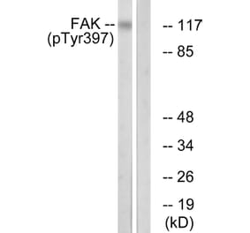
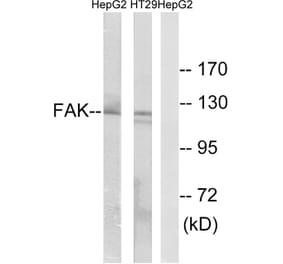
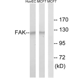
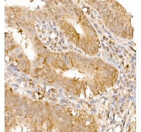
Tissue inhibitors of metalloproteinases (TIMPs) control extracellular matrix (ECM) homeostasis by inhibiting the activity of matrix metalloproteinases (MMPs), which are associated with ECM turnover. Recent studies have revealed that TIMPs are implicated in tumorigenesis in both MMP-dependent and MMP-independent manners. We examined a mechanism by which TIMP-2 stimulated lung adenocarcinoma cell proliferation, independent of MMP inhibition. The stimulation of growth by TIMP-2 in A549 cells required c-Src kinase activation. c-Src kinase activity, induced by TIMP-2, concomitantly increased FAK, phosphoinositide 3-kinase (PI3-kinase)/AKT, and ERK1/2 activation. Selective knockdown of integrin α3β1, known as a TIMP-2 receptor, did not significantly change TIMP-2 growth promoting activity. Furthermore, we showed that high TIMP-2 expression in lung adenocarcinomas is associated with a worse prognosis from multiple cohorts, especially for stage I lung adenocarcinoma. Through integrated analysis of The Cancer Genome Atlas data, TIMP-2 expression was significantly associated with the alteration of driving genes, c-Src activation, and PI3-kinase/AKT pathway activation. Taken together, our results demonstrate that TIMP-2 stimulates lung adenocarcinoma cell proliferation through c-Src, FAK, PI3-kinase/AKT, and ERK1/2 pathway activation in an MMP-independent manner.
The purpose of this study was to elucidate the molecular mechanisms of microRNA-205 (miR-205) as a tumor suppressor in prostate cancer (PCa). In the present study, microRNA microarray analysis suggested that the expression of miR-205 was significantly decreased in advanced PCa compared with early PCa. Real-time PCR analysis also indicated that miR-205 expression was significantly decreased in PCa tissues compared with non-cancerous tissues. Moreover, the expression of miR-205 has been demonstrated to be associated with the clinicopathological stage and total/free prostate-specific antigen (PSA) level of PCa. Functional analyses showed that both the overexpression of miR-205 and the knockdown of c-SRC in PCa cell lines could inhibit cell growth, colony formation, migration, invasion and the cell cycle as well as induce cell apoptosis in vitro. Furthermore, over-expressing miR-205 reduced tumorigenicity in vivo. Through a luciferase activity assay and Western blotting, c-SRC was identified as a target of miR-205 in cells. The overexpression of miR-205 suppressed c-SRC and its downstream signaling molecules, including FAK, p-FAK, ERK1/2 and p-ERK1/2, and attenuated cell proliferation, invasion and tumor growth.


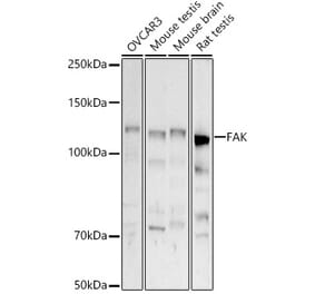
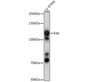
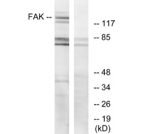
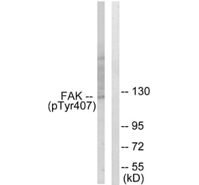
![Western Blot - Anti-FAK Antibody [ARC0171] (A13236) - Antibodies.com](https://cdn.antibodies.com/image/catalog/13/A13236_1.jpg?profile=product_alternative)