Primary Antibodies
Secondary Antibodies
Proteins & Peptides
ELISA Kits
About Us
Contact Us
Sign In/Register
0
ISO 9001:2015 Certified
Live Customer Support
4.5/5 on Trustpilot
100% Quality Guarantee
Home
Assistive Reagents
FAM alkyne, 5-isomer (A270204)
FAM alkyne, 5-isomer (A270204)
Overview
Specifications
Images
Enlarge Image
Enlarge Image
$110
Product Datasheet
100% Guarantee
Price Match Guarantee
Product Size:
1mg
5mg
10mg
25mg
50mg
100mg
Quantity:
1
2
3
4
5
6
7
8
9
10
Add To Cart
Request a Quotation
Custom or Bulk Request
Shipping Information
Freight/Packing Charges:
$40
Dispatched from St. Louis, MO.
Lead Time: 5-8 business days.
Specifications
Name
FAM alkyne, 5-isomer
Description
FAM (fluorescein) alkyne for copper-catalyzed Click chemistry, high purity (97+%) 5-isomer. The compound possesses significant aqueous solubility.
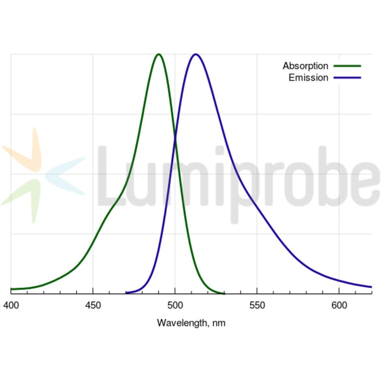
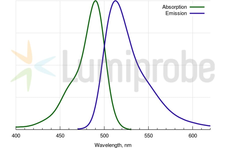
Absorption Maxima
490 nm
Extinction Coefficient
80000 M
-1
cm
-1
Emission Maxima
513 nm
Fluorescence Quantum Yield
0.93
CAS Number
510758-19-7
CF
260
0.20
CF
280
0.17
Purity
95% (by
1
H NMR and HPLC-MS).
Molecular Formula
C
25
H
15
O
6
N
Molecular Weight
413.38 Da
Product Form
Yellow solid.
Solubility
Good in aqueous buffers (pH > 8), alcohols, DMSO, and DMF.
Storage
Shipped at room temperature. Upon delivery, store in the dark at -20°C. Avoid prolonged exposure to light.
Disclaimer
This product is for research use only. It is not intended for diagnostic or therapeutic use.
Scientific Validation Data
Validation Data
(2)
Enlarge Image
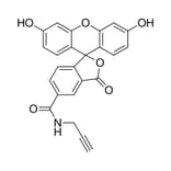
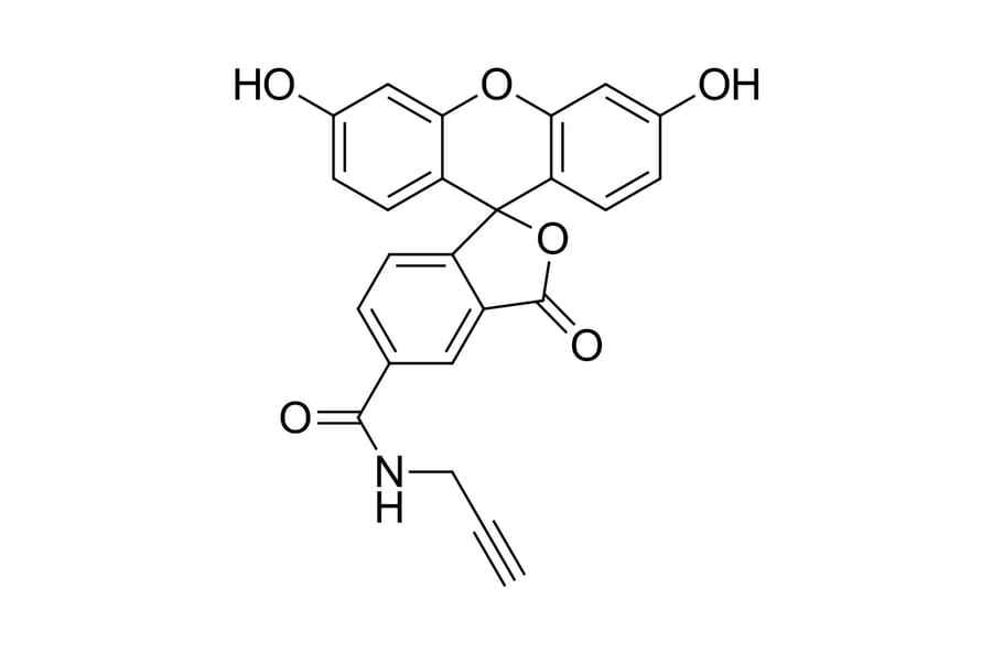
Chemical Structure - FAM alkyne, 5-isomer (A270204)
Structure of 5-FAM alkyne.
Enlarge Image
FAM alkyne, 5-isomer (A270204)
FAM absorbance and emission spectra.
Publishing research using FAM alkyne, 5-isomer (A270204)? Please
let us know
so that we can list the citation on this page.
Top