Primary Antibodies
Secondary Antibodies
Proteins & Peptides
ELISA Kits
About Us
Contact Us
Sign In/Register
0
ISO 9001:2015 Certified
Live Customer Support
4.5/5 on Trustpilot
100% Quality Guarantee
Home
Primary Antibodies
Fc epsilon RI alpha Antibodies
Anti-FCER1A Antibody (FITC) (A245)
Anti-FCER1A Antibody (FITC) (A245)
Overview
Specifications
Images
Enlarge Image
$590
Product Datasheet
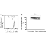
Mouse monoclonal (CRA1) antibody to FCER1A (FITC) for WB, FC, IHC-P, IHC-Fr and IF.
100% Guarantee
Price Match Guarantee
Product Size:
50µg
Quantity:
1
2
3
4
5
6
7
8
9
10
Add To Cart
Request a Quotation
Custom or Bulk Request
Shipping Information
Freight/Packing Charges:
$40
Dispatched from St. Louis, MO.
Lead Time: 4-6 business days.
Tags:
Basophil Marker
Mast Cell Marker
Specifications
Name
Anti-FCER1A Antibody (FITC)
Description
Mouse monoclonal (CRA1) antibody to FCER1A (FITC).
Applications
WB
, FC,
IHC-P
,
IHC-Fr
,
IF
Dilutions
IF: 2-10 µg/ml, FC: 2-10 µg/ml
Reactivity
Human
Immunogen
Recombinant extracellular portion of human FCER1A protein (corresponding to a.a. Met-26-197, where signal peptide is 1-25).
Epitope
Amino acids 26-110.
Host
Mouse
Clonality
Monoclonal
Clone ID
CRA1
Isotype
IgG2b
Conjugate
FITC
Excitation: 490nm, Emission: 525nm
Purification
Affinity purification.
Concentration
1 mg/ml
Product Form
Liquid
Formulation
Supplied in Phosphate Buffered Saline, pH 7.4, with 50% Glycerol (filter-sterilized, without Sodium Azide and carrier free).
Storage
Shipped at 4°C. Upon delivery aliquot and store at -20°C. Avoid freeze / thaw cycles.
Synonyms
Fc-epsilon RI-alpha, FCE1A, FcERI, High affinity immunoglobulin epsilon receptor subunit alpha, IgE Fc receptor subunit alpha
Isotype Controls
Mouse IgG2b [MPC-11] (FITC) (A86740)
Disclaimer
This product is for research use only. It is not intended for diagnostic or therapeutic use.
Scientific Validation Data
Validation Data
(1)
Enlarge Image
Anti-FCER1A Antibody (FITC) (A245)
Anti-FCER1A Antibody (FITC)
Publishing research using Anti-FCER1A Antibody (FITC) (A245)? Please
let us know
so that we can list the citation on this page.
Alternative products to Anti-FCER1A Antibody (FITC) (A245)
A246
Anti-FCER1A Antibody (FITC)
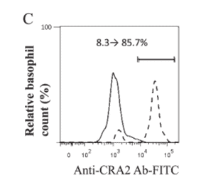
Mouse monoclonal (CRA2) antibody to FCER1A (FITC) for WB, IHC-Fr, IHC-P, ICC and FC.
A246
Anti-FCER1A Antibody (FITC)
Mouse monoclonal (CRA2) antibody to FCER1A (FITC) for WB, IHC-Fr, IHC-P, ICC and FC.
Proteins predicted to interact with Fc epsilon RI alpha
Predicted protein interactions based upon String database. Revelancy score correlates with probability of interaction.
FCER1G Antibodies
99.7% Relevancy Score
Syk Antibodies
96.9% Relevancy Score
Fyn Antibodies
94.7% Relevancy Score
CPA3 Antibodies
92.1% Relevancy Score
Lyn Antibodies
86% Relevancy Score
CD20 Antibodies
80.5% Relevancy Score
CD89 Antibodies
74.4% Relevancy Score
PAG1 Antibodies
72.7% Relevancy Score
c-Kit Antibodies
71.4% Relevancy Score
Top