Figure 1: Western Blot - Anti-Glucagon Antibody [ARC56934] (A309735)
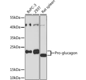
Western blot analysis of various lysates, using Anti-Glucagon Antibody [ARC56934] (A309735) at 1:10,000 dilution. The secondary antibody was Goat Anti-Rabbit IgG H&L Antibody (HRP) at 1:10,000 dilution. Lysates/proteins were present at 25µg per lane. The blocking buffer used was 3% non-fat dry milk in TBST. Detection was with a ECL Enhanced Kit (RM00021). Exposure time: 180s.
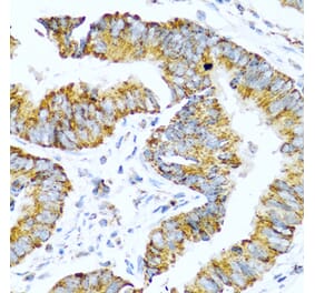
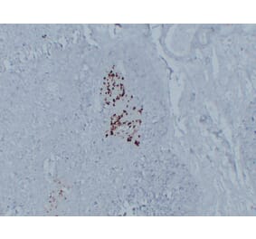
Immunohistochemistry analysis of paraffin-embedded human pancreas using Anti-Glucagon Antibody [ARC56934] (A309735) at a dilution of 1:50000/100000 (40x lens). Perform high pressure antigen retrieval with 10 mM Tris/EDTA buffer pH 9.0 before commencing with IHC staining protocol.
Confocal imaging of human pancreas using Anti-Glucagon Antibody [ARC56934] (A309735), at a dilution of 1:1000, (red). DAPI was used for nuclear staining (Blue). Objective: 60x.
Confocal imaging of mouse pancreas using Anti-Glucagon Antibody [ARC56934] (A309735), at a dilution of 1:1000, (red). DAPI was used for nuclear staining (Blue). Objective: 60x.
Confocal imaging of rat pancreas using Anti-Glucagon Antibody [ARC56934] (A309735), at a dilution of 1:1000, (red). DAPI was used for nuclear staining (Blue). Objective: 60x.


![Western Blot - Anti-Glucagon Antibody [ARC56934] (A309735) - Antibodies.com](https://cdn.antibodies.com/image/catalog/309/A309735_1.jpg?profile=product_top)
![Immunohistochemistry - Anti-Glucagon Antibody [ARC56934] (A309735) - Antibodies.com](https://cdn.antibodies.com/image/catalog/309/A309735_2.jpg?profile=product_top)
![Immunofluorescence - Anti-Glucagon Antibody [ARC56934] (A309735) - Antibodies.com](https://cdn.antibodies.com/image/catalog/309/A309735_3.jpg?profile=product_top)
![Immunofluorescence - Anti-Glucagon Antibody [ARC56934] (A309735) - Antibodies.com](https://cdn.antibodies.com/image/catalog/309/A309735_4.jpg?profile=product_top)
![Immunofluorescence - Anti-Glucagon Antibody [ARC56934] (A309735) - Antibodies.com](https://cdn.antibodies.com/image/catalog/309/A309735_5.jpg?profile=product_top)
![Western Blot - Anti-Glucagon Antibody [ARC56934] (A309735) - Antibodies.com](https://cdn.antibodies.com/image/catalog/309/A309735_1.jpg?profile=product_top_thumb)
![Immunohistochemistry - Anti-Glucagon Antibody [ARC56934] (A309735) - Antibodies.com](https://cdn.antibodies.com/image/catalog/309/A309735_2.jpg?profile=product_top_thumb)
![Immunofluorescence - Anti-Glucagon Antibody [ARC56934] (A309735) - Antibodies.com](https://cdn.antibodies.com/image/catalog/309/A309735_3.jpg?profile=product_top_thumb)
![Immunofluorescence - Anti-Glucagon Antibody [ARC56934] (A309735) - Antibodies.com](https://cdn.antibodies.com/image/catalog/309/A309735_4.jpg?profile=product_top_thumb)
![Immunofluorescence - Anti-Glucagon Antibody [ARC56934] (A309735) - Antibodies.com](https://cdn.antibodies.com/image/catalog/309/A309735_5.jpg?profile=product_top_thumb)
![Western Blot - Anti-Glucagon Antibody [ARC56934] (A309735) - Antibodies.com](https://cdn.antibodies.com/image/catalog/309/A309735_1.jpg?profile=product_image)
![Immunohistochemistry - Anti-Glucagon Antibody [ARC56934] (A309735) - Antibodies.com](https://cdn.antibodies.com/image/catalog/309/A309735_2.jpg?profile=product_image)
![Immunofluorescence - Anti-Glucagon Antibody [ARC56934] (A309735) - Antibodies.com](https://cdn.antibodies.com/image/catalog/309/A309735_3.jpg?profile=product_image)
![Immunofluorescence - Anti-Glucagon Antibody [ARC56934] (A309735) - Antibodies.com](https://cdn.antibodies.com/image/catalog/309/A309735_4.jpg?profile=product_image)
![Immunofluorescence - Anti-Glucagon Antibody [ARC56934] (A309735) - Antibodies.com](https://cdn.antibodies.com/image/catalog/309/A309735_5.jpg?profile=product_image)
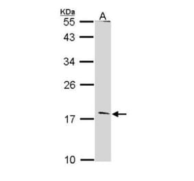
![Western Blot - Anti-Glucagon Antibody [ARC0382] (A309737) - Antibodies.com](https://cdn.antibodies.com/image/catalog/309/A309737_1.jpg?profile=product_alternative)

![Immunohistochemistry - Anti-Glucagon Antibody [ARC56936] (A307477) - Antibodies.com](https://cdn.antibodies.com/image/catalog/307/A307477_1.jpg?profile=product_alternative)
![Immunofluorescence - Anti-Glucagon Antibody [ARC1143] (A308918) - Antibodies.com](https://cdn.antibodies.com/image/catalog/308/A308918_1.jpg?profile=product_alternative)