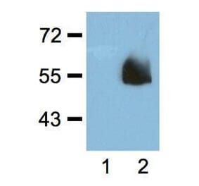
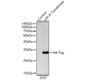
Figure 1: Western Blot - Anti-HA Tag Antibody [ARC59578] (A309245)
Western blot analysis of 293F-NFAT, using Anti-HA Tag Antibody [ARC59578] (A309245) at 1:10,000 dilution. Lysates/proteins were present at 25µg per lane. The blocking buffer used was 3% non-fat dry milk in TBST. Detection was with a ECL Basic Kit. Exposure time: 1s.
Figure 2: Immunofluorescence - Anti-HA Tag Antibody [ARC59578] (A309245)
Immunofluorescence analysis of 293T-SCRNI-HA-GFP(C) and 293T cells using Anti-HA Tag Antibody [ARC59578] (A309245) at a dilution of 1:100 (40x lens). DAPI was used to stain the cell nuclei (blue).
Figure 3: Immunofluorescence - Anti-HA Tag Antibody [ARC59578] (A309245)
Immunofluorescence analysis of 293T-SCRNI-HA-GFP(N) and 293T cells using Anti-HA Tag Antibody [ARC59578] (A309245) at a dilution of 1:100 (40x lens). DAPI was used to stain the cell nuclei (blue).
Publishing research using Anti-HA Tag Antibody [ARC59578] (A309245)? Please let us know so that we can list the citation on this page.
Alternative products to Anti-HA Tag Antibody [ARC59578] (A309245)


![Western Blot - Anti-HA Tag Antibody [ARC59578] (A309245) - Antibodies.com](https://cdn.antibodies.com/image/catalog/309/A309245_1.jpg?profile=product_top)
![Immunofluorescence - Anti-HA Tag Antibody [ARC59578] (A309245) - Antibodies.com](https://cdn.antibodies.com/image/catalog/309/A309245_2.jpg?profile=product_top)
![Immunofluorescence - Anti-HA Tag Antibody [ARC59578] (A309245) - Antibodies.com](https://cdn.antibodies.com/image/catalog/309/A309245_3.jpg?profile=product_top)
![Western Blot - Anti-HA Tag Antibody [ARC59578] (A309245) - Antibodies.com](https://cdn.antibodies.com/image/catalog/309/A309245_1.jpg?profile=product_top_thumb)
![Immunofluorescence - Anti-HA Tag Antibody [ARC59578] (A309245) - Antibodies.com](https://cdn.antibodies.com/image/catalog/309/A309245_2.jpg?profile=product_top_thumb)
![Immunofluorescence - Anti-HA Tag Antibody [ARC59578] (A309245) - Antibodies.com](https://cdn.antibodies.com/image/catalog/309/A309245_3.jpg?profile=product_top_thumb)
![Western Blot - Anti-HA Tag Antibody [ARC59578] (A309245) - Antibodies.com](https://cdn.antibodies.com/image/catalog/309/A309245_1.jpg?profile=product_image)
![Immunofluorescence - Anti-HA Tag Antibody [ARC59578] (A309245) - Antibodies.com](https://cdn.antibodies.com/image/catalog/309/A309245_2.jpg?profile=product_image)
![Immunofluorescence - Anti-HA Tag Antibody [ARC59578] (A309245) - Antibodies.com](https://cdn.antibodies.com/image/catalog/309/A309245_3.jpg?profile=product_image)
![SDS-PAGE - Anti-HA Tag Antibody [HA/279] - BSA and Azide free (A278453) - Antibodies.com](https://cdn.antibodies.com/image/catalog/278/A278453_1.jpg?profile=product_alternative)
![Western Blot - Anti-HA Tag Antibody [16.43] - BSA and Azide free (A254042) - Antibodies.com](https://cdn.antibodies.com/image/catalog/254/A254043_1.jpg?profile=product_alternative)
![Western Blot - Anti-HA Tag Antibody [RM305] (A121321) - Antibodies.com](https://cdn.antibodies.com/image/catalog/121/A121321_1.png?profile=product_alternative)
![Western Blot - Anti-HA Tag Antibody [16.43] (A250862) - Antibodies.com](https://cdn.antibodies.com/image/catalog/250/A250863_1.jpg?profile=product_alternative)
![SDS-PAGE - Anti-HA Tag Antibody [HA/279] (A277865) - Antibodies.com](https://cdn.antibodies.com/image/catalog/277/A277865_1.jpg?profile=product_alternative)
![ELISA - Anti-HA Tag Antibody [RMH02] (A121359) - Antibodies.com](https://cdn.antibodies.com/image/catalog/121/A121322_1.png?profile=product_alternative)