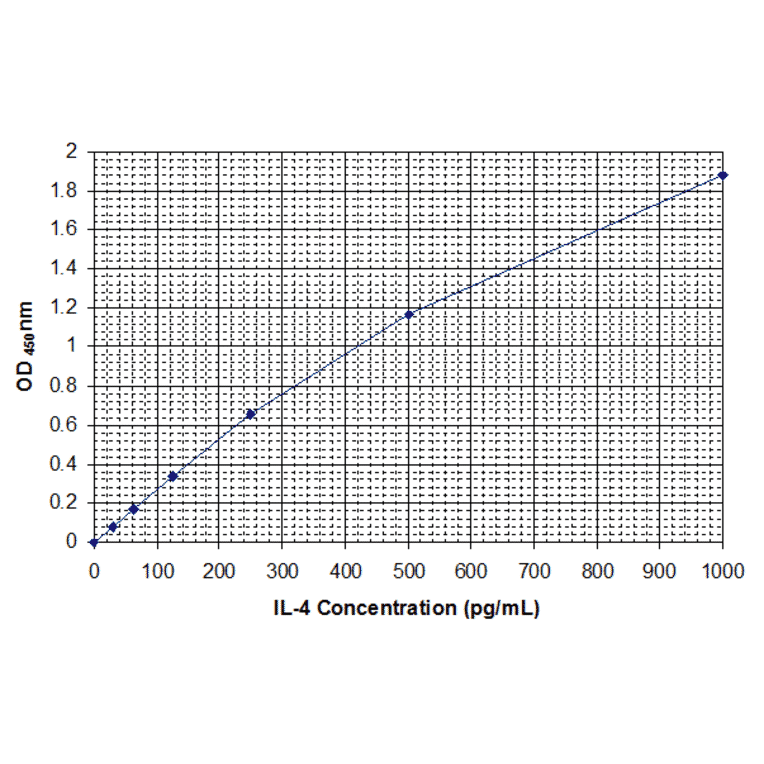
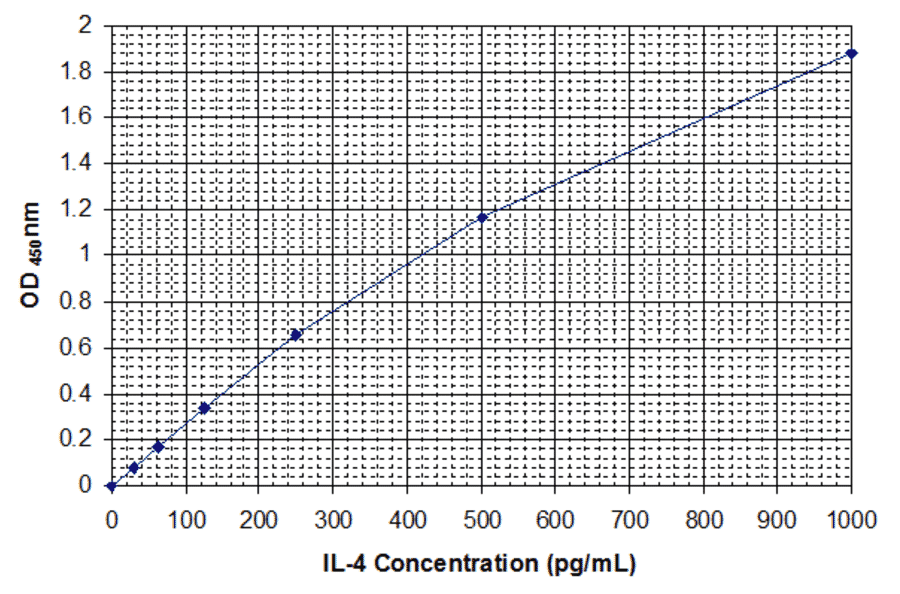
The ectoenzyme CD39 hydrolyzes extracellular adenosine 5'-triphosphate (ATP), which possesses pro-inflammatory properties. However, the role of CD39 in allergic asthma has not been fully elucidated. A total of 18 patients with persistent asthma who were allergic to house dust mites and 19 healthy volunteers were enrolled in this study. The expression of CD39, GATA3, RAR-related orphan receptor γ (ROR-γt) and forkhead box P3 (FoxP3) mRNA in peripheral blood mononuclear cells (PBMCs) was determined by SYBR-Green I quantitative polymerase chain reaction (qPCR). The cytokines interleukin (IL)-4, IL-17A, transforming growth factor β (TGF-β) and DP.sIgE were detected by enzyme-linked immunosorbent assay. Our data demonstrated that the expression of CD39 mRNA in PBMCs from asthmatic patients was significantly lower compared to that in normal controls [(1.49±0.59)×10-3 vs. (2.17±0.77)×10-3, respectively; P<0.01]. CD39 mRNA was negatively correlated with serum IL-4, IL-17A and GATA3 expression (r=-0.468, P<0.05; r=-0.550, P<0.05; and r=-0.424, P<0.01, respectively) and positively correlated with FoxP3 and TGF-β expression (r=0.373, P<0.05; and r=0.425, P<0.05, respectively). There was no obvious correlation between CD39 and ROR-γt expression (r=-0.259, P=0.122). These data suggested that CD39 mRNA expression was downregulated in allergic asthma, which was positively correlated with serum IL-4, IL-17A and GATA3 expression and negatively correlated with serum TGF-β and FoxP3 expression, whereas there was no correlation with ROR-γt. Therefore, it was hypothesized that CD39 may participate in the occurrence and progression of allergic asthma.
OBJECTIVE:
The aim of the present research was to verify the levels of the soluble adhesion molecules sL- and sE-selectins, intercellular adhesion molecule (sICAM)-1, and vascular cell adhesion molecule-1 in serial samples of internal jugular venous blood taken from migraine patients without aura (MWoA) during attacks. The expression of leukocyte function antigen (LFA)-1 and very late activation antigen (VLA)-4 was also assessed on lymphocytes obtained from jugular venous blood. Levels of certain proinflammatory cytokines (tumor necrosis factor-alpha[TNF-alpha], interleukin-1beta[IL-1beta], IL-4, and IL-6) were also determined and correlated with those of adhesion molecules.
PATIENTS AND METHODS:
Seven MWoA patients were admitted in the hospital during attacks and blood samples were taken immediately after catheter insertion, at 1, 2, and 4 hours after attack onset, and within 2 hours after its termination. The levels of adhesion molecules and cytokines were measured with ELISA method. The expression of LFA-1 and VLA-4 was assessed by flow cytometry.
RESULTS:
A parallel transient increase of sICAM-1, TNF-alpha, and IL-6 was observed in the first 2 hours after attack onset compared with the time of catheter insertion (P < .0001, <.001, and <.003, respectively). The proportion of CD4+ and CD8+ T-cells expressing high levels of LFA-1 showed instead a progressive down-regulation with significantly lower percentages at 2 and 4 hours after attack onset (P < .01 and <.022, respectively). No variation in the percentage of VLA-4 expressing cells was observed at any time of the study.
CONCLUSIONS:
The transient increase in sICAM-1 and TNF-alpha found in the internal jugular blood of MWoA patients assessed ictally can be induced by sensory neuropeptides released from activated trigeminal endings. The progressive decrease in sICAM-1 levels during attacks and the down-regulation of LFA-1 expression by lymphocytes could antagonize their transvascular migration, supporting the hypothesis of sterile inflammation in the dura mater during migraine attacks.