

Unconjugated
Breast cancer is a major cause of cancer-related death in women. Antitumor T cell responses play critical therapeutic roles, including direct cytotoxicity mediated by CD8+ T cells and immunomodulatory roles mediated by CD4+ T cells. The IL-9-expressing Th9 cells are recently found to present antitumor immunity in melanoma and lung adenocarcinoma. In this study, we found that IL-9 expression in the serum and in circulating CD4+ T cells were significantly upregulated in breast cancer patients compared to healthy controls. The IL-9-expressing Th9 cells were enriched in the CCR4-CCR6-CXCR3- subset. Upon TCR stimulation, this subset also presented potent IL-10 and IL-21 expression in addition to IL-9 expression. CCR4-CCR6-CXCR3- CD4+ T cells also assisted in the killing of autologous tumor cells by CD8+ T cells, but did not initiate cytotoxicity by themselves. This enhancement in CD8+ T cell-mediated cytotoxicity was dependent on IL-9 as well as on IL-21. Interestingly, the tumor-infiltrating Th9 cells presented comparable IL-9, reduced IL-10, and elevated IL-21 expression compared with their counterparts in the peripheral blood. Together, these results demonstrated that IL-9-expressing Th9 cells were upregulated in breast cancer patients and potentially possessed antitumor roles by enhancing CD8+ T cell-mediated cytotoxicity.
Interleukin (IL)-21 is crucial for the regulation of lymphocytes and is implicated in autoimmune and other diseases. The relevance of being able to measure human IL-21 prompted us to develop ELISA and ELISpot assays for analysis of IL-21 levels and IL-21-producing cells, respectively. Monoclonal antibodies (mAbs) to IL-21 were made and ELISA and ELISpot assays were developed. The selected detection mAb also neutralized IL-21-mediated activation of human cells. Peripheral blood mononuclear cells (PBMCs) from healthy donors (n=24) were stimulated polyclonally (phytohemagglutinin; PHA) or with antigen (Candida albicans extract and tetanus toxoid). Using ELISpot, high numbers of IL-21-producing cells were detected after PHA activation; lower but positive responses to antigen were seen in approximately 50% of the donors. In contrast, the ELISA detected IL-21 in supernatants from PHA-activated cells but not from antigen-stimulated cells. When analyzing IL-17A in parallel, PHA and antigens induced detectable responses in ELISpot as well as in ELISA. Hypothesizing that the lack of detectable IL-21 levels after antigenic stimulation was due to a combination of low frequencies of IL-21-secreting cells and consumption of IL-21 by cellular receptors during cell culture, PBMCs (n=18) were stimulated in the presence of the neutralizing detection mAb. When preventing IL-21 from interacting with its receptor, increased IL-21 levels were found by ELISA after PHA activation and IL-21 could also be measured after antigen stimulation. ELISpot results were unaffected by the addition of the neutralizing mAb. In conclusion, IL-21 secreted by low frequencies of antigen-specific ex vivo-stimulated PBMC can be difficult to detect by ELISA but prevention of IL-21 interaction with its receptor leads to detectable IL-21 levels. In ELISpot, where the cytokine is captured by mAbs on a solid phase immediately upon secretion, blocking the receptor interaction does not affect the detection of IL-21-secreting cells.


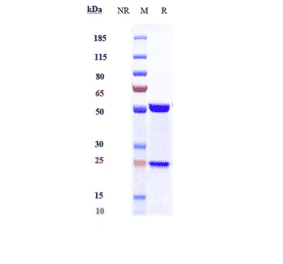
![SDS-PAGE - Anti-IL-21 Antibody [Research Grade Biosimilar] - Low endotoxin, Azide free (A324088) - Antibodies.com](https://cdn.antibodies.com/image/catalog/324/A324088_1.jpg?profile=product_alternative)
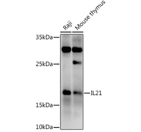
![Western Blot - Anti-IL-21 Antibody [ARC60383] (A309923) - Antibodies.com](https://cdn.antibodies.com/image/catalog/309/A309923_1.jpg?profile=product_alternative)
![ELISA - Anti-IL-21 Humanized Antibody [Avizakimab Biosimilar] - Azide free (A318862) - Antibodies.com](https://cdn.antibodies.com/image/catalog/318/A318862_1.jpg?profile=product_alternative)