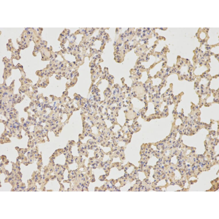
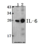
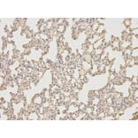
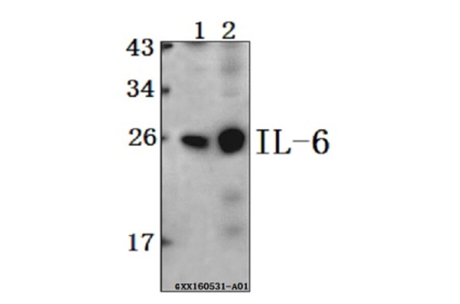
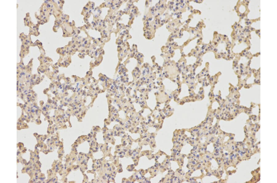
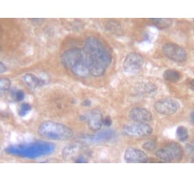
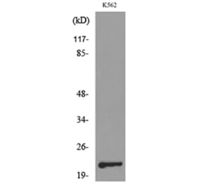
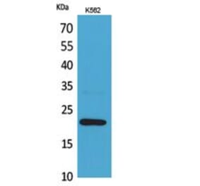
IL-6 pAb detects endogenous levels of IL-6 protein.
Applications
WB, IHC
Reactivity
Human, Mouse, Rat
Immunogen
Recombinant full length Human IL-6.
Host
Rabbit
Clonality
Polyclonal
Conjugate
Unconjugated
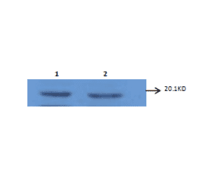
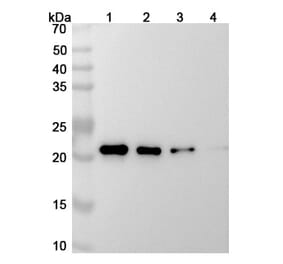
Molecular Weight
~ 23 kDa
Purity
The antibody was affinity-purified from rabbit antiserum by affinity-chromatography using epitope-specific immunogen and the purity is > 95% (by SDS-PAGE).
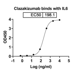
Recombinant humanized monoclonal antibody to IL-6 for use as a research grade Clazakizumab biosimila for ELISA, Flow Cytometry, Functional Studies and in vivo Research.
Recombinant humanized monoclonal antibody to IL-6 for use as a research grade Olokizumab biosimilar for ELISA, Flow Cytometry, Functional Studies and in vivo Research.
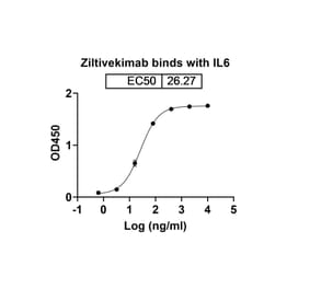
Recombinant humanized monoclonal antibody to IL-6 for use as a research grade Ziltivekimab biosimila for ELISA, Flow Cytometry, Functional Studies and in vivo Research.
Recombinant chimeric monoclonal antibody to IL-6 for use as a research grade Siltuximab biosimilar for ELISA, Flow Cytometry, Functional Studies and in vivo Research.
Recombinant humanized monoclonal antibody to IL-6 for use as a research grade Clazakizumab biosimila for ELISA, Flow Cytometry, Functional Studies and in vivo Research.
Recombinant humanized monoclonal antibody to IL-6 for use as a research grade Olokizumab biosimilar for ELISA, Flow Cytometry, Functional Studies and in vivo Research.
Recombinant humanized monoclonal antibody to IL-6 for use as a research grade Ziltivekimab biosimila for ELISA, Flow Cytometry, Functional Studies and in vivo Research.
Recombinant chimeric monoclonal antibody to IL-6 for use as a research grade Siltuximab biosimilar for ELISA, Flow Cytometry, Functional Studies and in vivo Research.


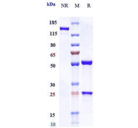
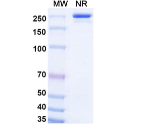
![SDS-PAGE - Anti-IL-6 Antibody [Research Grade Biosimilar] - Low endotoxin, Azide free (A324098) - Antibodies.com](https://cdn.antibodies.com/image/catalog/324/A324098_1.jpg?profile=product_alternative)
![SDS-PAGE - Anti-IL-6 Antibody [MEDI 5117] - Low endotoxin, Azide free (A324100) - Antibodies.com](https://cdn.antibodies.com/image/catalog/324/A324100_1.jpg?profile=product_alternative)
![SDS-PAGE - Anti-IL-6 Antibody [Research Grade Biosimilar] - Low endotoxin, Azide free (A324096) - Antibodies.com](https://cdn.antibodies.com/image/catalog/324/A324096_1.jpg?profile=product_alternative)
![SDS-PAGE - Anti-IL-6 Antibody [Research Grade Biosimilar] - Low endotoxin, Azide free (A324097) - Antibodies.com](https://cdn.antibodies.com/image/catalog/324/A324097_1.jpg?profile=product_alternative)