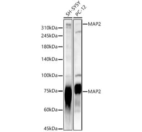
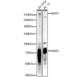
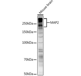
Figure 1: Western Blot - Anti-MAP2 Antibody [ARC56285] (A306845)
Western blot analysis of Rat brain, using Anti-MAP2 Antibody [ARC56285] (A306845) at 1:4,000 dilution. The secondary antibody was Goat Anti-Rabbit IgG H&L Antibody (HRP) at 1:10,000 dilution. Lysates/proteins were present at 25µg per lane. The blocking buffer used was 3% non-fat dry milk in TBST. Detection was with a ECL Enhanced Kit (RM00021). Exposure time: 180s.
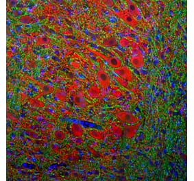
Immunohistochemistry analysis of paraffin-embedded human brain tissue using Anti-MAP2 Antibody [ARC56285] (A306845) at a dilution of 1:200(40x lens). Perform high pressure antigen retrieval with 10 mM citrate buffer pH 6.0 before commencing with IHC staining protocol.
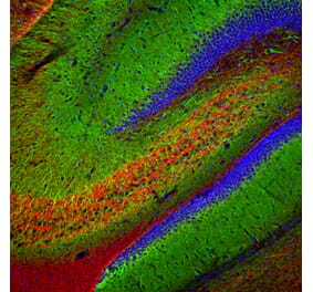
Immunohistochemistry analysis of paraffin-embedded mouse brain using Anti-MAP2 Antibody [ARC56285] (A306845) at a dilution of 1:200(40x lens). Perform high pressure antigen retrieval with 10 mM citrate buffer pH 6.0 before commencing with IHC staining protocol.
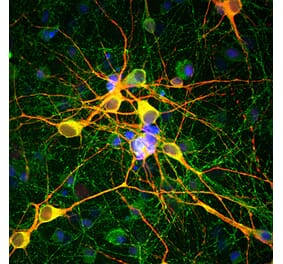
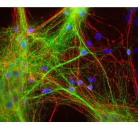
Confocal imaging of SH-SYSY cells using Anti-MAP2 Antibody [ARC56285] (A306845), at a dilution of 1:200, (red). The cells were counterstained with Anti-alpha Tubulin Antibody, at a dilution of 1:400, (green). DAPI was used for nuclear staining (Blue). Objective: 60x.
Publishing research using Anti-MAP2 Antibody [ARC56285] (A306845)? Please let us know so that we can list the citation on this page.
Alternative products to Anti-MAP2 Antibody [ARC56285] (A306845)


![Western Blot - Anti-MAP2 Antibody [ARC56285] (A306845) - Antibodies.com](https://cdn.antibodies.com/image/catalog/306/A306845_1.jpg?profile=product_top)
![Immunohistochemistry - Anti-MAP2 Antibody [ARC56285] (A306845) - Antibodies.com](https://cdn.antibodies.com/image/catalog/306/A306845_2.jpg?profile=product_top)
![Immunohistochemistry - Anti-MAP2 Antibody [ARC56285] (A306845) - Antibodies.com](https://cdn.antibodies.com/image/catalog/306/A306845_3.jpg?profile=product_top)
![Immunofluorescence - Anti-MAP2 Antibody [ARC56285] (A306845) - Antibodies.com](https://cdn.antibodies.com/image/catalog/306/A306845_4.jpg?profile=product_top)
![Western Blot - Anti-MAP2 Antibody [ARC56285] (A306845) - Antibodies.com](https://cdn.antibodies.com/image/catalog/306/A306845_1.jpg?profile=product_top_thumb)
![Immunohistochemistry - Anti-MAP2 Antibody [ARC56285] (A306845) - Antibodies.com](https://cdn.antibodies.com/image/catalog/306/A306845_2.jpg?profile=product_top_thumb)
![Immunohistochemistry - Anti-MAP2 Antibody [ARC56285] (A306845) - Antibodies.com](https://cdn.antibodies.com/image/catalog/306/A306845_3.jpg?profile=product_top_thumb)
![Immunofluorescence - Anti-MAP2 Antibody [ARC56285] (A306845) - Antibodies.com](https://cdn.antibodies.com/image/catalog/306/A306845_4.jpg?profile=product_top_thumb)
![Western Blot - Anti-MAP2 Antibody [ARC56285] (A306845) - Antibodies.com](https://cdn.antibodies.com/image/catalog/306/A306845_1.jpg?profile=product_image)
![Immunohistochemistry - Anti-MAP2 Antibody [ARC56285] (A306845) - Antibodies.com](https://cdn.antibodies.com/image/catalog/306/A306845_2.jpg?profile=product_image)
![Immunohistochemistry - Anti-MAP2 Antibody [ARC56285] (A306845) - Antibodies.com](https://cdn.antibodies.com/image/catalog/306/A306845_3.jpg?profile=product_image)
![Immunofluorescence - Anti-MAP2 Antibody [ARC56285] (A306845) - Antibodies.com](https://cdn.antibodies.com/image/catalog/306/A306845_4.jpg?profile=product_image)
![Immunofluorescence - Anti-MAP2 Antibody [2C4] (A85459) - Antibodies.com](https://cdn.antibodies.com/image/catalog/85/A85459_1.jpg?profile=product_alternative)
![Western Blot - Anti-MAP2 Antibody [MT-07] (A86616) - Antibodies.com](https://cdn.antibodies.com/image/catalog/86/A86618_762.jpg?profile=product_alternative)
![Western Blot - Anti-MAP2 Antibody [MT-08] (A86618) - Antibodies.com](https://cdn.antibodies.com/image/catalog/86/A86619_763.jpg?profile=product_alternative)
![Western Blot - Anti-MAP2 Antibody [ARC56279] (A306846) - Antibodies.com](https://cdn.antibodies.com/image/catalog/306/A306846_1.jpg?profile=product_alternative)
![Western Blot - Anti-MAP2 Antibody [MT-01] (A86785) - Antibodies.com](https://cdn.antibodies.com/image/catalog/86/A86786_893.jpg?profile=product_alternative)
