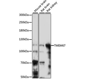
Figure 1: Western Blot - Anti-Meckelin Antibody [ARC3051] (A309404)
Western blot analysis of extracts of various cell lines, using Anti-Meckelin Antibody [ARC3051] (A309404) at 1:1,000 dilution. The secondary antibody was Goat Anti-Rabbit IgG H&L Antibody (HRP) at 1:10,000 dilution. Lysates/proteins were present at 25µg per lane. The blocking buffer used was 3% non-fat dry milk in TBST. Detection was with a ECL Basic Kit. Exposure time: 90s.
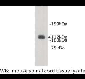
Figure 2: Western Blot - Anti-Meckelin Antibody [ARC3051] (A309404)
Western blot analysis of extracts of PC-12 cells, using Anti-Meckelin Antibody [ARC3051] (A309404) at 1:1,000 dilution. The secondary antibody was Goat Anti-Rabbit IgG H&L Antibody (HRP) at 1:10,000 dilution. Lysates/proteins were present at 25µg per lane. The blocking buffer used was 3% non-fat dry milk in TBST. Detection was with a ECL Enhanced Kit (RM00021). Exposure time: 90s.
Immunohistochemistry analysis of paraffin-embedded human colon carcinoma tissue using Anti-Meckelin Antibody [ARC3051] (A309404) at a dilution of 1:200(40x lens). Perform high pressure antigen retrieval with 10 mM citrate buffer pH 6.0 before commencing with IHC staining protocol.
Immunohistochemistry analysis of paraffin-embedded mouse kidney using Anti-Meckelin Antibody [ARC3051] (A309404) at a dilution of 1:200(40x lens). Perform high pressure antigen retrieval with 10 mM citrate buffer pH 6.0 before commencing with IHC staining protocol.
Immunohistochemistry analysis of paraffin-embedded rat liver using Anti-Meckelin Antibody [ARC3051] (A309404) at a dilution of 1:200(40x lens). Perform high pressure antigen retrieval with 10 mM citrate buffer pH 6.0 before commencing with IHC staining protocol.


![Western Blot - Anti-Meckelin Antibody [ARC3051] (A309404) - Antibodies.com](https://cdn.antibodies.com/image/catalog/309/A309404_1.jpg?profile=product_top)
![Western Blot - Anti-Meckelin Antibody [ARC3051] (A309404) - Antibodies.com](https://cdn.antibodies.com/image/catalog/309/A309404_2.jpg?profile=product_top)
![Immunohistochemistry - Anti-Meckelin Antibody [ARC3051] (A309404) - Antibodies.com](https://cdn.antibodies.com/image/catalog/309/A309404_3.jpg?profile=product_top)
![Immunohistochemistry - Anti-Meckelin Antibody [ARC3051] (A309404) - Antibodies.com](https://cdn.antibodies.com/image/catalog/309/A309404_4.jpg?profile=product_top)
![Immunohistochemistry - Anti-Meckelin Antibody [ARC3051] (A309404) - Antibodies.com](https://cdn.antibodies.com/image/catalog/309/A309404_5.jpg?profile=product_top)
![Western Blot - Anti-Meckelin Antibody [ARC3051] (A309404) - Antibodies.com](https://cdn.antibodies.com/image/catalog/309/A309404_1.jpg?profile=product_top_thumb)
![Western Blot - Anti-Meckelin Antibody [ARC3051] (A309404) - Antibodies.com](https://cdn.antibodies.com/image/catalog/309/A309404_2.jpg?profile=product_top_thumb)
![Immunohistochemistry - Anti-Meckelin Antibody [ARC3051] (A309404) - Antibodies.com](https://cdn.antibodies.com/image/catalog/309/A309404_3.jpg?profile=product_top_thumb)
![Immunohistochemistry - Anti-Meckelin Antibody [ARC3051] (A309404) - Antibodies.com](https://cdn.antibodies.com/image/catalog/309/A309404_4.jpg?profile=product_top_thumb)
![Immunohistochemistry - Anti-Meckelin Antibody [ARC3051] (A309404) - Antibodies.com](https://cdn.antibodies.com/image/catalog/309/A309404_5.jpg?profile=product_top_thumb)
![Western Blot - Anti-Meckelin Antibody [ARC3051] (A309404) - Antibodies.com](https://cdn.antibodies.com/image/catalog/309/A309404_1.jpg?profile=product_image)
![Western Blot - Anti-Meckelin Antibody [ARC3051] (A309404) - Antibodies.com](https://cdn.antibodies.com/image/catalog/309/A309404_2.jpg?profile=product_image)
![Immunohistochemistry - Anti-Meckelin Antibody [ARC3051] (A309404) - Antibodies.com](https://cdn.antibodies.com/image/catalog/309/A309404_3.jpg?profile=product_image)
![Immunohistochemistry - Anti-Meckelin Antibody [ARC3051] (A309404) - Antibodies.com](https://cdn.antibodies.com/image/catalog/309/A309404_4.jpg?profile=product_image)
![Immunohistochemistry - Anti-Meckelin Antibody [ARC3051] (A309404) - Antibodies.com](https://cdn.antibodies.com/image/catalog/309/A309404_5.jpg?profile=product_image)