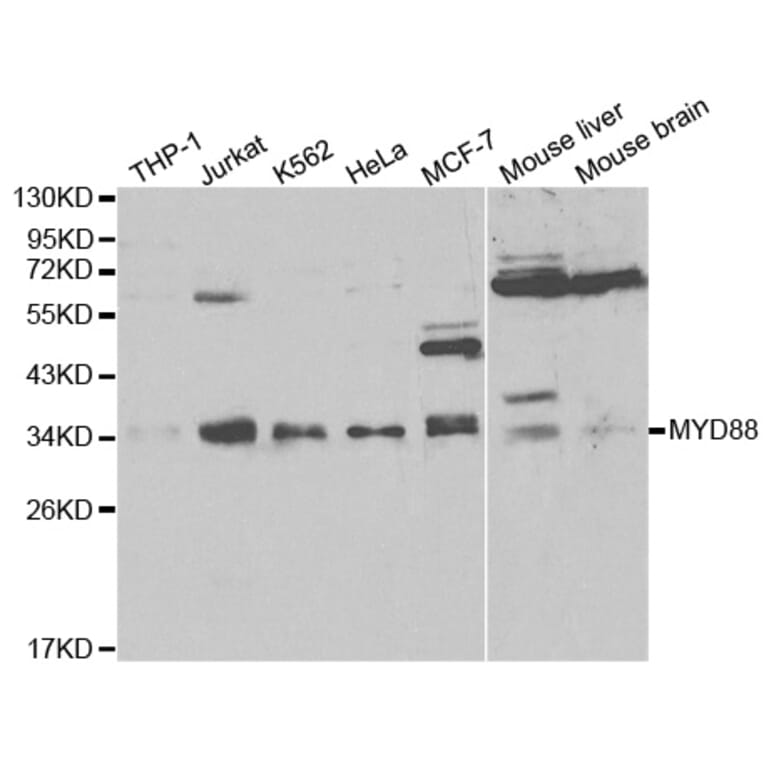
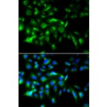
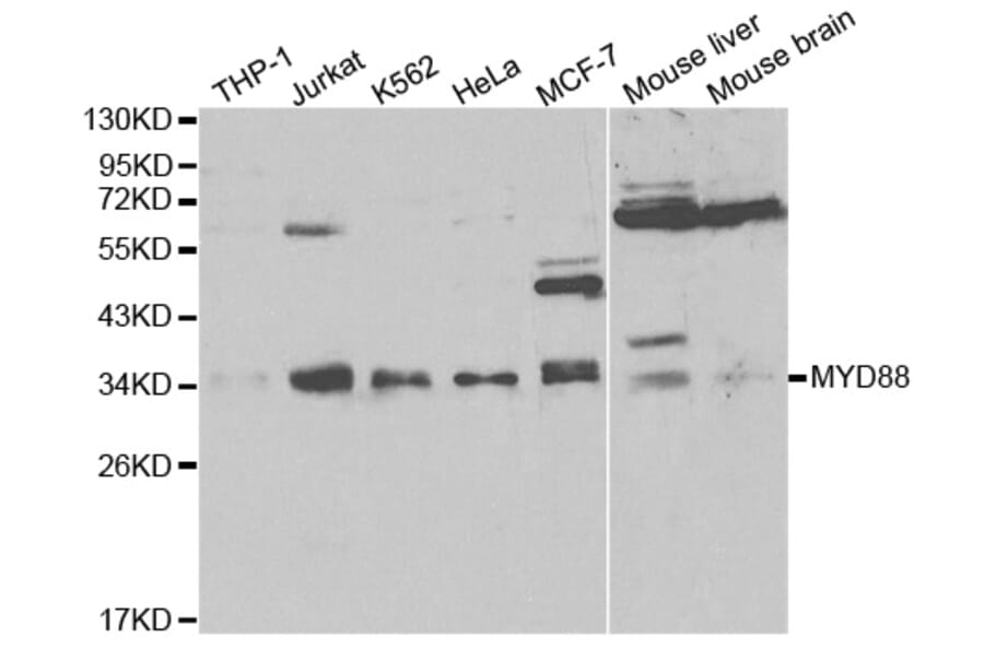
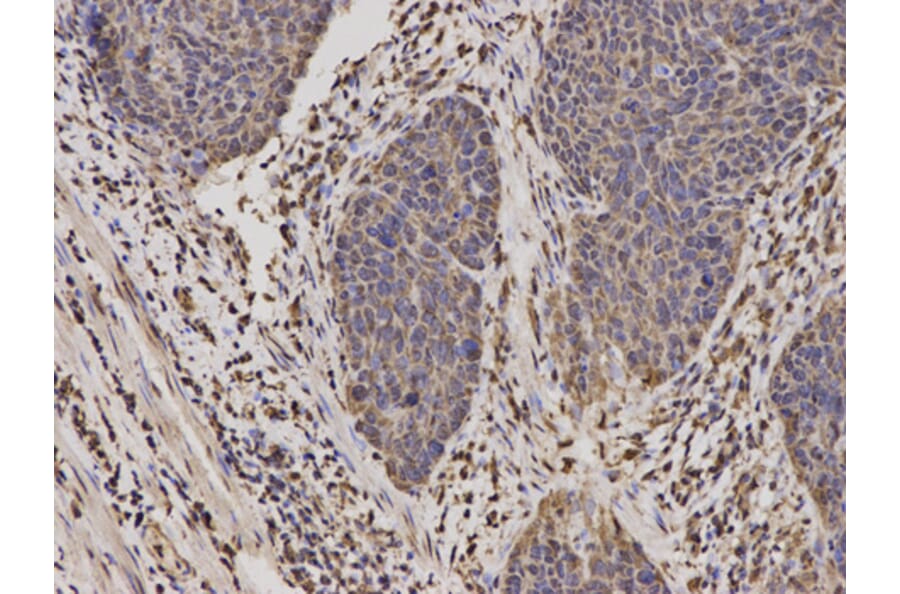
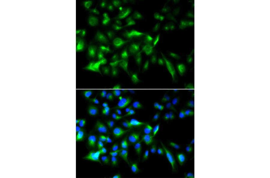
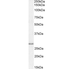
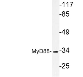
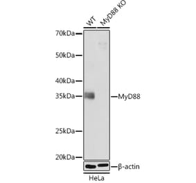
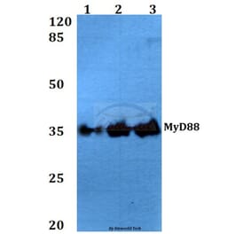
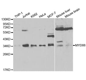
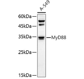
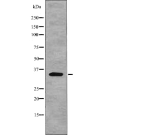
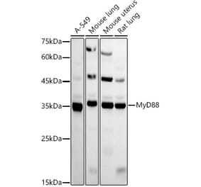
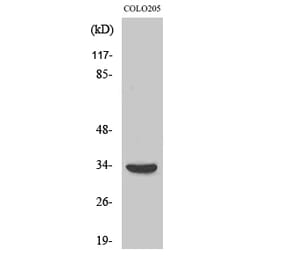
MYD88 pAb detects endogenous levels of MYD88 protein.
Applications
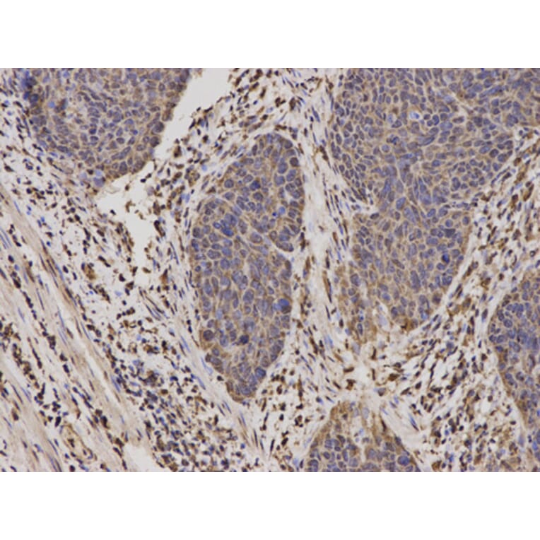
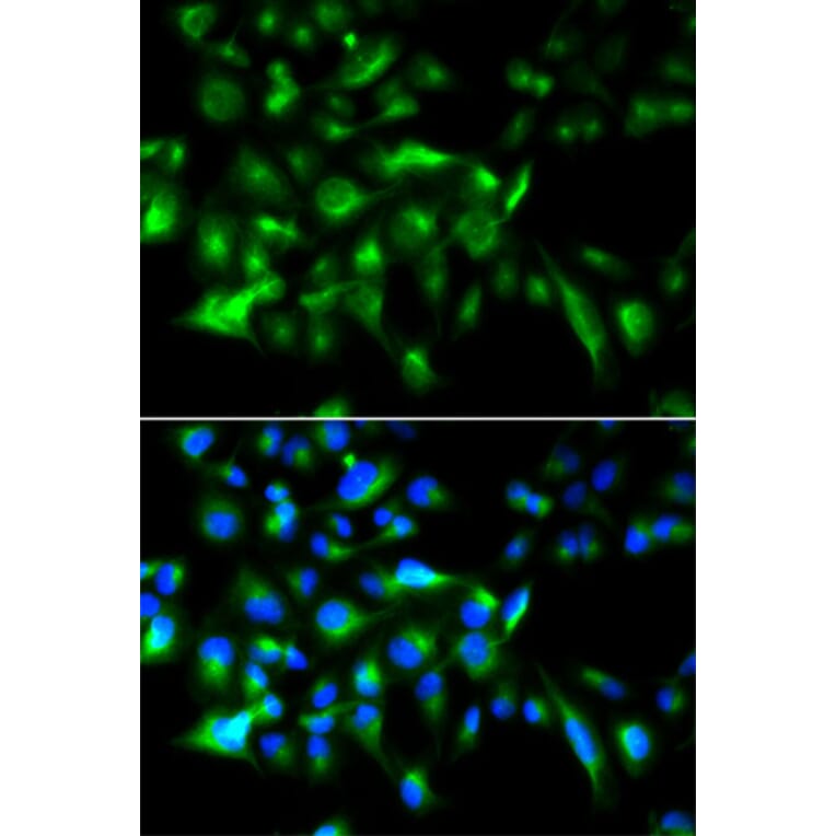
WB, IHC, IF
Reactivity
Human, Mouse, Rat
Immunogen
Recombinant full length Human MyD88.
Host
Rabbit
Clonality
Polyclonal
Conjugate
Unconjugated
Molecular Weight
~ 35 kDa
Purity
The antibody was affinity-purified from rabbit antiserum by affinity-chromatography using epitope-specific immunogen and the purity is > 95% (by SDS-PAGE).
Product Form
1mg/ml in PBSwith0.1%SodiumAzide,50%Glycerol.
Synonyms
Myeloid differentiation primary response protein MyD88