Anti-NFkB-p65 (phospho-S536) Antibody (A27418) has been discontinued and is no longer available.
View all available products.
Unconjugated
Vascular endothelial dysfunction is regarded as the initial step of vascular complications in diabetes mellitus. This study investigated the amelioration of apigenin and naringenin in type 2 diabetic (T2D) rats induced by high-fat diet and streptozotocin and explored the underlying mechanism. Apigenin or naringenin was intragastrically administered at 50 or 100mg/kg once a day for 6 weeks. Biochemical parameters including blood glucose, glycated serum protein, serum lipid, insulin, superoxide dismutase (SOD), malonaldehyde and intercellular adhesion molecule-1 (ICAM-1) were measured. Vascular reactivity in isolated thoracic aortic rings was examined. Pathological features of the thoracic aorta were further observed through optical microscopy and transmission electron microscopy. Lastly, we evaluated their effects on insulin resistance of palmitic acid (PA)-induced endothelial cells. Compared with diabetic control group, apigenin and naringenin significantly decreased the levels of blood glucose, serum lipid, malonaldehyde, ICAM-1 and insulin resistance index, increased SOD activity and improved impaired glucose tolerance. Apigenin and naringenin restored phenylephrine-mediated contractions and acetylcholine or insulin-induced relaxations in aortic tissues. Furthermore, pathological damage in the thoracic aorta of apigenin and naringenin groups was more remissive than diabetic control group. In vitro, apigenin and naringenin inhibited NF-κB activation and ICAM-1 mRNA expression in PA-treated endothelial cells and improved nitric oxide production in the presence of insulin. In conclusion, both apigenin and naringenin can ameliorate glucose and lipid metabolism, as well as endothelial dysfunction in T2D rats at least in part by down-regulating oxidative stress and inflammation. In general, apigenin showed greater potency than naringenin equivalent.
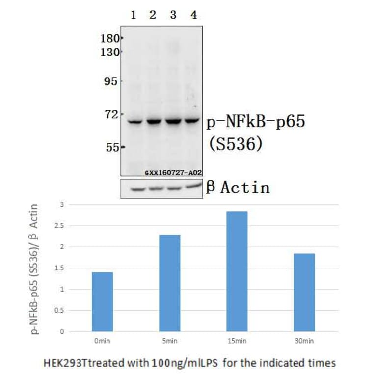
Endotoxin can stimulate inflammatory cytokine release from monocytes/macrophages and result in septic shock. Glycyrrhetinic acid (GA), the main bioactive component of licorice, possesses substantial anti-inflammatory activity. Here, we explored effect of 11-deoxy-18α-glycyrrhetinic acid-30-ethyl ester (DGAEE), a newly synthesized derivative of GA, on septic shock. DGAEE and its main metabolite 11-deoxy-18α-glycyrrhetinic acid (DGA) significantly alleviated septic shock as evidenced by improvements of survival rates, lung histopathological changes and wet/dry ratio in lipopolysaccharide (LPS)/D-galactosamine-stimulated mice, and decreased blood pressure in LPS/D-galactosamine-stimulated rats. The two compounds decreased serum levels of NO, TNF-α, IL-6, IL-1β, and increased the level of IL-10 more potently in mice. In LPS-stimulated RAW 264.7 cells, DGA but not DGAEE showed marked regulation of NO, TNF-α, IL-6 and IL-10 levels, suggesting that DGAEE display anti-shock effect by DGA rather than itself. Moreover, the neutralizing antibody against IL-10 markedly prohibited the inhibitory effect of DGA on the production of cytokines from RAW 264.7 cells, and AS101 (an inhibitor of IL-10 biosynthesis) almost completely reversed the anti-shock effect of DGA in mice. In addition, DGA did not affect activation of NF-κB-p65 and p38 MAPK as well as IκBα degradation, but moderately reduced activation of ERK and JNK, and markedly increased phosphorylation of GSK3β in LPS-stimulated RAW 264.7 cells. LY294002 (an inhibitor of GSK3β phosphorylation) and LiCl (an inhibitor of GSK3β activity) diminished and potentiated increase of IL-10 levels by DGA, respectively. In conclusion, DGAEE alleviates septic shock through DGA in an IL-10-dependent manner, and the mechanism is related to inactivation of GSK3β.