Unconjugated
Metformin, the most widely administered oral anti‑diabetic therapeutic agent, exerts its glucose-lowering effect predominantly via liver kinase B1 (LKB1)-dependent activation of adenosine monophosphate-activated protein kinase (AMPK). Accumulating evidence has demonstrated that metformin possesses potential antitumor effects. However, whether the antitumor effect of metformin is via the LKB1/AMPK signaling pathway remains to be determined. In the current study, the effects of metformin on proliferation, cell cycle progression, and apoptosis of human non‑small cell lung cancer (NSCLC) H460 (LKB1‑null) and H1299 (LKB1‑positive) cells were assessed, and the role of LKB1/AMPK signaling in the anti‑growth effects of metformin were investigated. Cell viability was determined using a 3-(4,5-dimethylthiazol-2-yl)-2,5-diphenyltetrazolium bromide assay, cell cycle distribution and apoptosis were assessed by flow cytometry, and protein expression levels were measured by western blotting. Metformin inhibited proliferation, induced significant cell cycle arrest at the G0‑G1 phase and increased apoptosis in NSCLC cells in a time- and concentration-dependent manner, regardless of the level of LKB1 protein expression. Furthermore, knockdown of LKB1 with short hairpin RNA (shRNA) did not affect the antiproliferative effect of metformin in the H1299 cells. Metformin stimulated AMPK phosphorylation and subsequently suppressed the phosphorylation of mammalian target of rapamycin and its downstream effector, 70‑kDa ribosomal protein S6 kinase in the two cell lines. These effects were abrogated by silencing AMPK with small interfering RNA (siRNA). In addition, knockdown of AMPK with siRNA inhibited the effect of metformin on cell proliferation in the two cell lines. These results provide evidence that the growth inhibition of metformin in NSCLC cells is mediated by LKB1‑independent activation of AMPK, indicating that metformin may be a potential therapeutic agent for the treatment of human NSCLC.
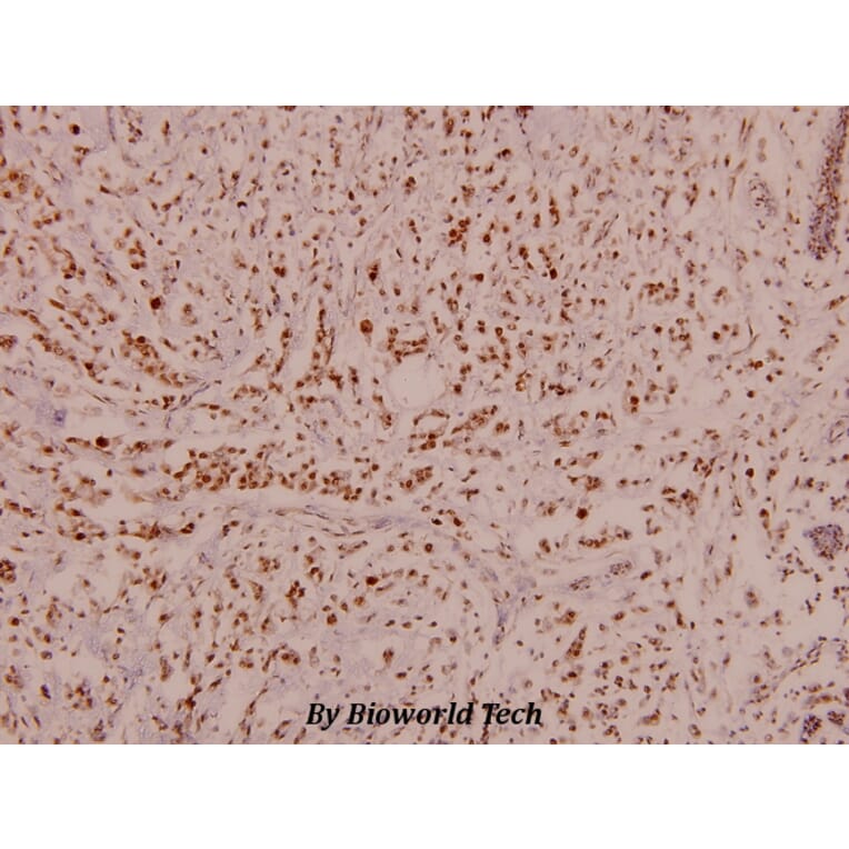
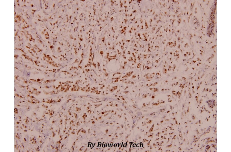
Autophagy plays a protective role in colorectal carcinoma. Arginine ADP-ribosyltransferase 1 (ART1) is an important mono-ADP-ribose transferase, which has been shown to play a role in biological processes such as proliferation and invasion of cancer cells. Interestingly, the role of ART1 in the regulation of autophagy is still not clear. We examined effects of overexpression or knockdown of ART1 by lentiviral transfection on starvation-induced autophagy of colon carcinoma CT26 cell lines in vivo and in vitro. The formation of autophagosome was detected by electron microscopy, acridine orange staining and expression of LC3 B. The molecular contributions of ART1 in regulation of autophagy were detected by western blotting or by co-immunoprecipitation. Additionally, inhibitors were used to study further the signaling pathway of ART1 in the regulation of autophagy. CCK8 assay, plate cloning assay, soft agar assay, examination of subcutaneous transplanted carcinoma in BALB/c mice, flow cytometry and Hoechst33342 staining were used to assess survival and apoptotic ability when starvation-induced autophagy modulated by ART1 was inhibited by 3-MA. Overexpression of ART1 promoted starvation-induced autophagy, which related to increases in the expression of Rac1, NF-κB, PARP-1, LKB1 and p-AMPK and a decrease in the expression of p-P70S6K. Correspondingly, knockdown of ART1 caused the opposite effects. ART1 also interacted with integrin α7. Additionally, changes of protein expressions were further validated following inhibition of Rac1 and PARP-1 in the starvation-induced ART1-GFP CT26 cells. Inhibition of ART1-stimulated starvation-induced autophagy restrained the growth and promoted apoptosis. ART1 is thus relevant in starvation-induced autophagy in colorectal carcinoma and may play essential roles in therapeutic anticancer strategies.