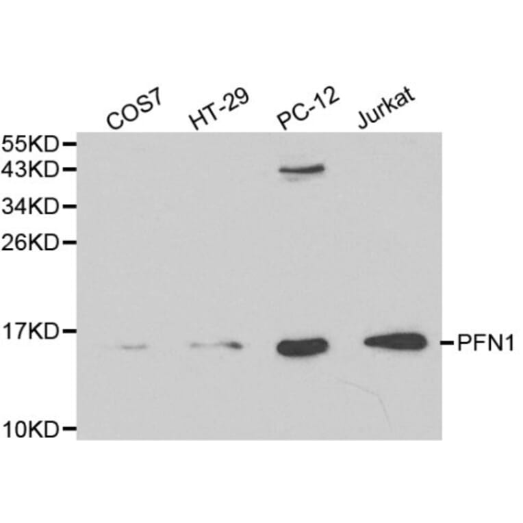
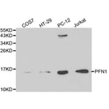
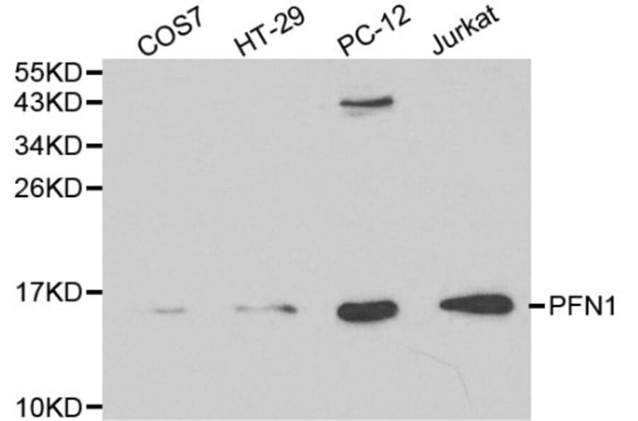
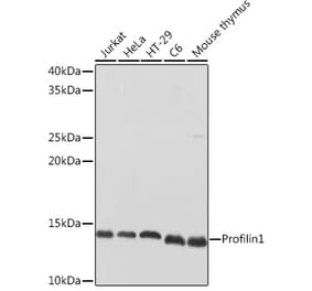
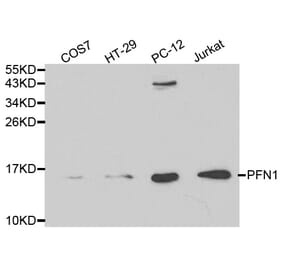
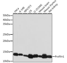
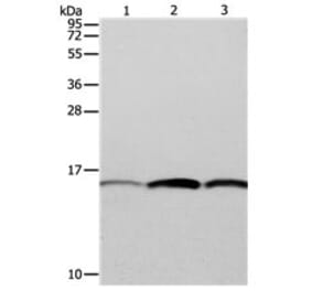
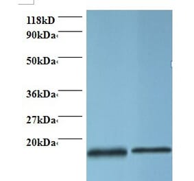
Unconjugated
BACKGROUND:
Neuroinflammation occurs in insulted regions of the brain and may be due to reactive oxygen species (ROS), nitric oxide (NO), cytokines, and chemokines produced by activated glia. Excessive production of neurotoxic molecules causes further neuronal damage. Low levels of vitamin D3 are a risk factor for various brain diseases.
METHODS:
Using the bacterial endotoxin, lipopolysaccharide (LPS), to induce neuroinflammation in primary cortical neuron-glia cultures, we investigated how 1,25-dihydroxyvitamin D3 (1,25(OH)2D3) affected neuroinflammation.
RESULTS:
LPS (100 ng/ml) induced the accumulation of nitrite and the production of ROS, interleukin (IL)-6, and macrophage inflammatory protein (MIP)-2 in time-dependent manners. Inhibition of p38 and extracellular signal-regulated kinase (ERK) but not c-Jun N-terminal kinase (JNK) mitogen-activated protein kinase (MAPK) by 20 μM of SB203580, PD98059, and SP600125, significantly reduced LPS-induced ROS production, NO accumulation, and inducible NO synthase (iNOS) expression, respectively. LPS-induced IL-6 and MIP-2 were significantly attenuated by inhibition of p38, ERK, and JNK MAPK. Cotreatment with 1,25(OH)2D3 attenuated LPS-induced ROS production, NO accumulation, and iNOS expression in concentration-dependent manners. 1,25(OH)2D3 also reduced LPS-induced production of IL-6 and MIP-2. Similarly, iNOS, IL-6, and MIP-2 mRNA expression in cells treated with LPS significantly increased, whereas this effect was attenuated by 1,25(OH)2D3. Moreover, LPS-induced phosphorylation of p38, ERK, and JNK MAPK was significantly inhibited by 1,25(OH)2D3.
CONCLUSIONS:
Our findings indicate that 1,25(OH)2D3 reduced the LPS-stimulated production of inflammatory molecules in neuron-glia cultures by inhibiting MAPK pathways and the production of downstream inflammatory molecules. We suggest that 1,25(OH)2D3 can be used to alleviate neuroinflammation in various brain injuries.
INTRODUCTION:
Acquired tamoxifen resistance remains the major obstacle to breast cancer endocrine therapy. β1-integrin was identified as one of the target genes of G protein-coupled estrogen receptor (GPER), a novel estrogen receptor recognized as an initiator of tamoxifen resistance. Here, we investigated the role of β1-integrin in GPER-mediated tamoxifen resistance in breast cancer.
METHODS:
The expression of β1-integrin and biomarkers of epithelial-mesenchymal transition were evaluated immunohistochemically in 53 specimens of metastases and paired primary tumors. The function of β1-integrin was investigated in tamoxifen-resistant (MCF-7R) subclones, derived from parental MCF-7 cells, and MCF-7R β1-integrin-silenced subclones in MTT and Transwell assays. Involved signaling pathways were identified using specific inhibitors and Western blotting analysis.
RESULTS:
GPER, β1-integrin and mesenchymal biomarkers (vimentin and fibronectin) expression in metastases increased compared to the corresponding primary tumors; a close expression pattern of β1-integrin and GPER were in metastases. Increased β1-integrin expression was also confirmed in MCF-7R cells compared with MCF-7 cells. This upregulation of β1-integrin was induced by agonists of GPER and blocked by both antagonist and knockdown of it in MCF-7R cells. Moreover, the epidermal growth factor receptor/extracellular regulated protein kinase (EGFR/ERK) signaling pathway was involved in this transcriptional regulation since specific inhibitors of these kinases also reduced the GPER-induced upregulation of β1-integrin. Interestingly, silencing of β1-integrin partially rescued the sensitivity of MCF-7R cells to tamoxifen and the α5β1-integrin subunit is probably responsible for this phenomenon. Importantly, the cell migration and epithelial-mesenchymal transition induced by cancer-associated fibroblasts, or the product of cancer-associated fibroblasts, fibronectin, were reduced by knockdown of β1-integrin in MCF-7R cells. In addition, the downstream kinases of β1-integrin including focal adhesion kinase, Src and AKT were activated in MCF-7R cells and may be involved in the interaction between cancer cells and cancer-associated fibroblasts.
CONCLUSIONS:
GPER/EGFR/ERK signaling upregulates β1-integrin expression and activates downstream kinases, which contributes to cancer-associated fibroblast-induced cell migration and epithelial-mesenchymal transition, in MCF-7R cells. GPER probably contributes to tamoxifen resistance via interaction with the tumor microenvironment in a β1-integrin-dependent pattern. Thus, β1-integrin may be a potential target to improve anti-hormone therapy responses in breast cancer patients.