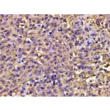
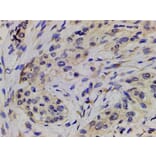
AF3241 at 1/200 staining human meningeal carcinomatosis(MC) tissue sections by IHC-P. The tissue was formaldehyde fixed and a heat mediated antigen retrieval step in citrate buffer was performed. The tissue was then blocked and incubated with the antibody for 1.5 hours at 22°C. An HRP conjugated goat anti-rabbit antibody was used as the secondary antibody.
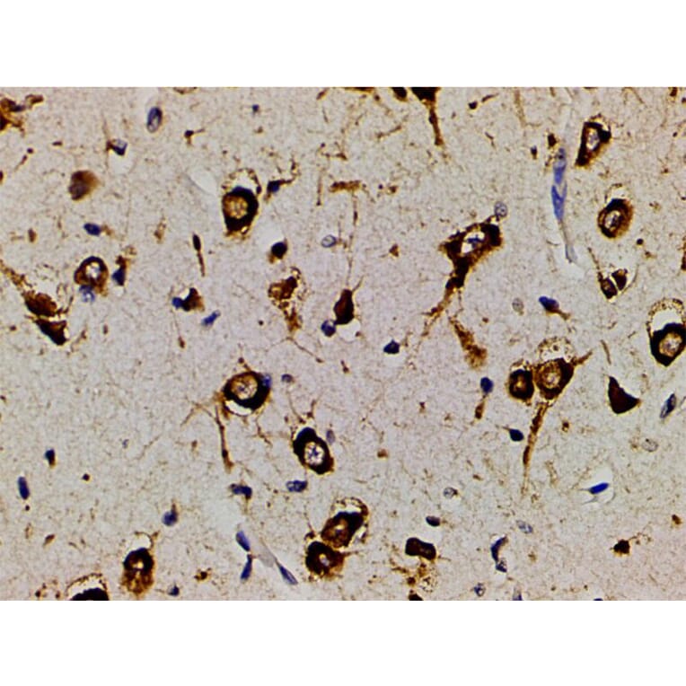
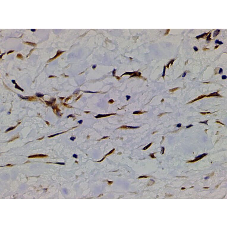
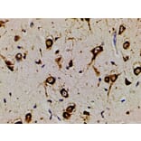
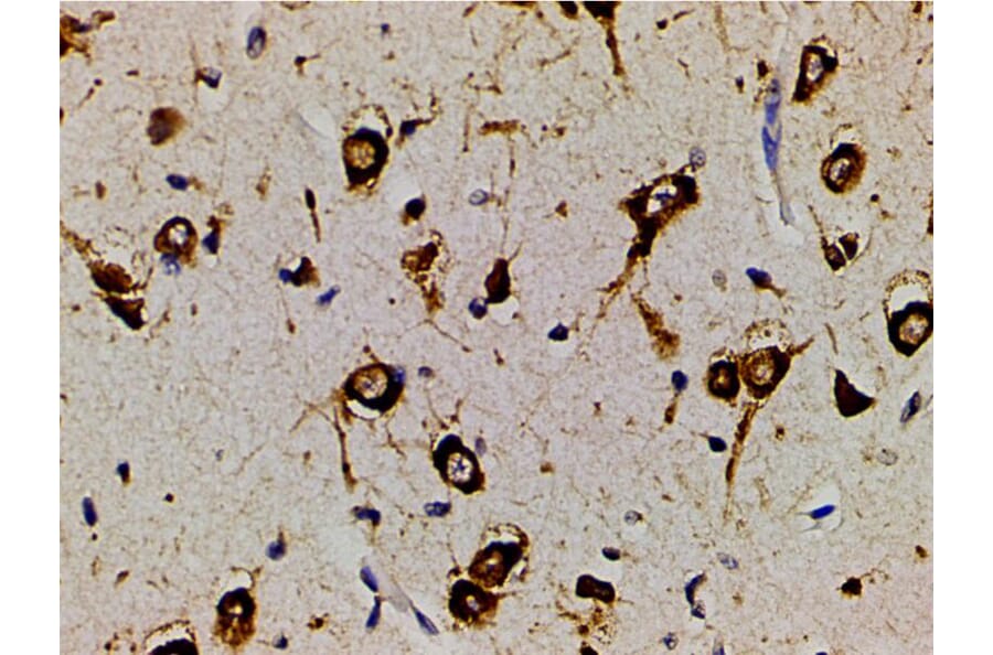
AF3241 at 1/200 staining human brain tissue sections by IHC-P. The tissue was formaldehyde fixed and a heat mediated antigen retrieval step in citrate buffer was performed. The tissue was then blocked and incubated with the antibody for 1.5 hours at 22°C. An HRP conjugated goat anti-rabbit antibody was used as the secondary antibody.
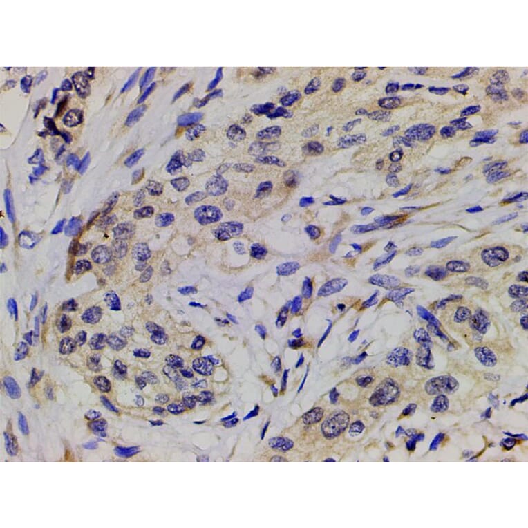
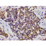
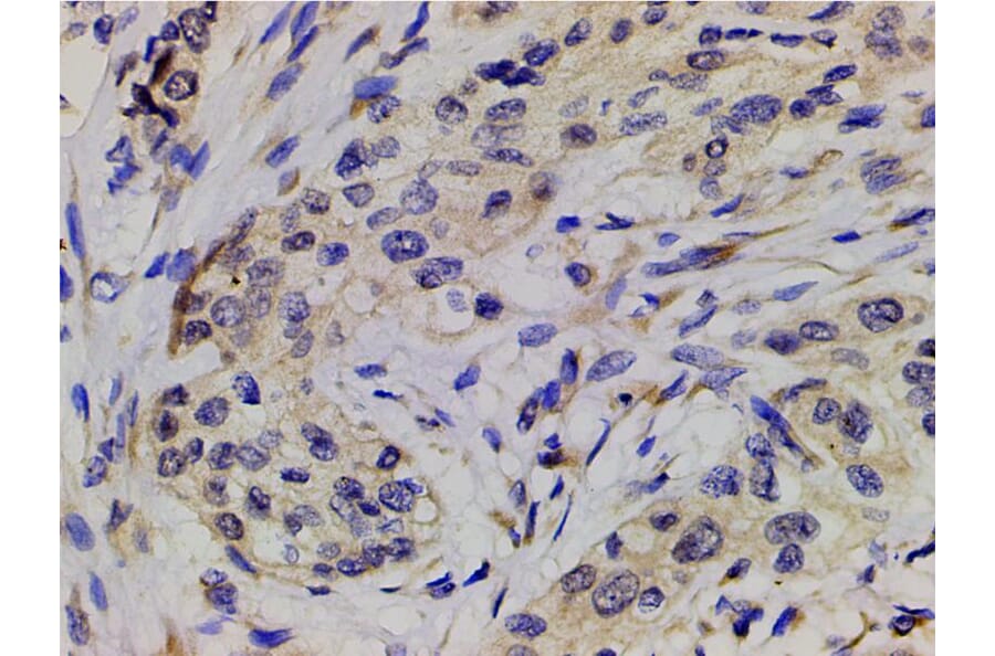
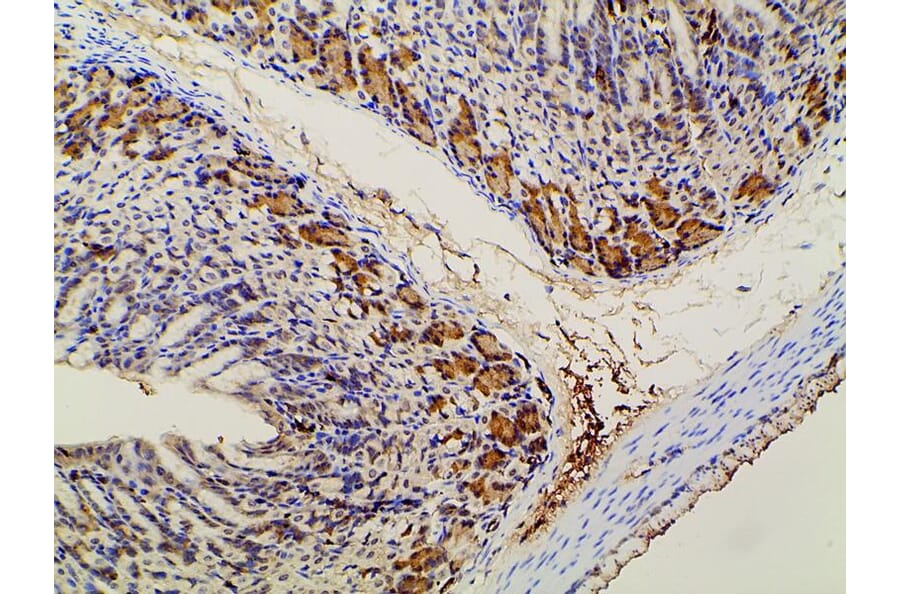
AF3241 at 1/200 staining human esophageal carcinoma tissue sections by IHC-P. The tissue was formaldehyde fixed and a heat mediated antigen retrieval step in citrate buffer was performed. The tissue was then blocked and incubated with the antibody for 1.5 hours at 22°C. An HRP conjugated goat anti-rabbit antibody was used as the secondary antibody.
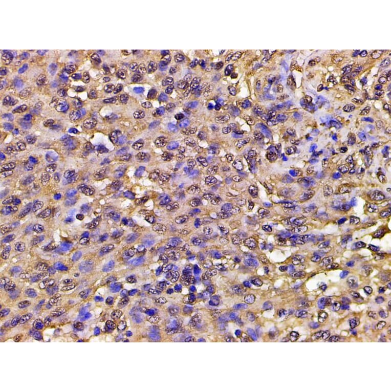
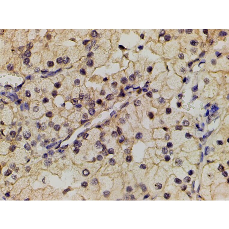
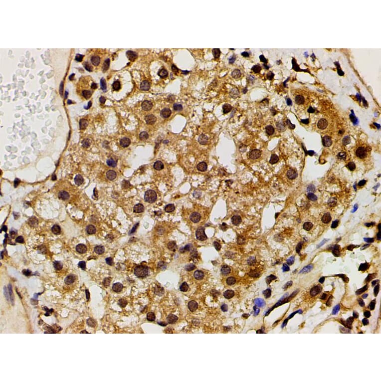
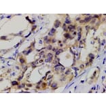
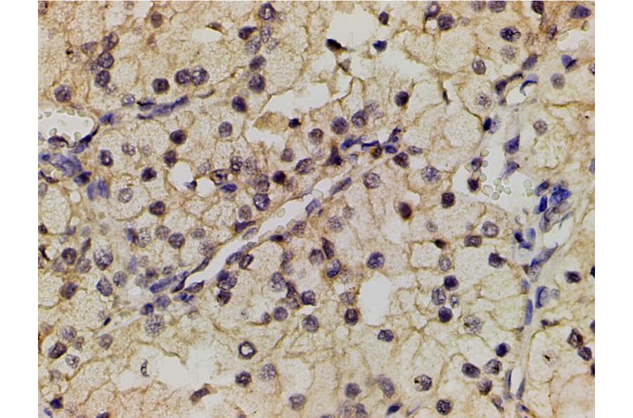
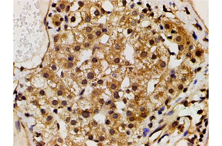
AF3241 at 1/200 staining human liver cancer tissue sections by IHC-P. The tissue was formaldehyde fixed and a heat mediated antigen retrieval step in citrate buffer was performed. The tissue was then blocked and incubated with the antibody for 1.5 hours at 22°C. An HRP conjugated goat anti-rabbit antibody was used as the secondary antibody.
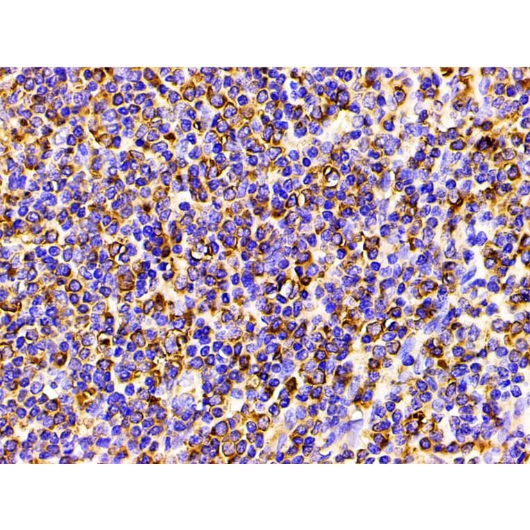
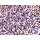
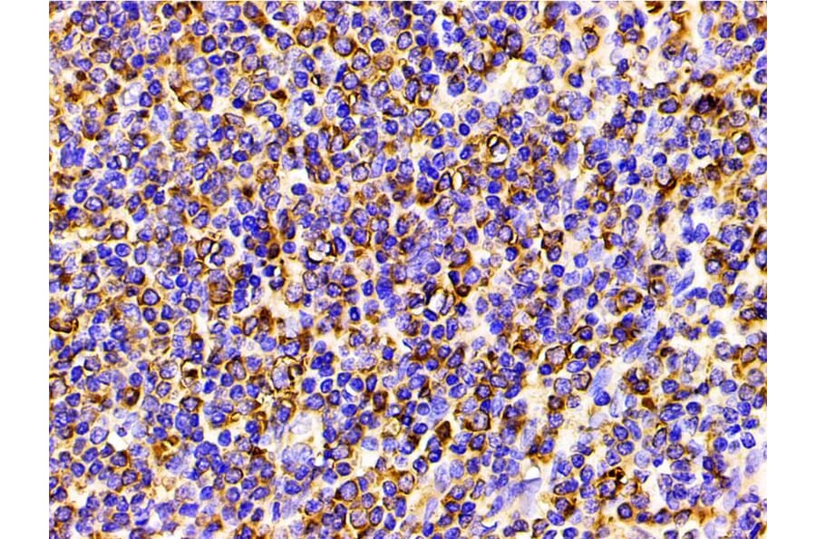
AF3241 at 1/200 staining human spleen tissue sections by IHC-P. The tissue was formaldehyde fixed and a heat mediated antigen retrieval step in citrate buffer was performed. The tissue was then blocked and incubated with the antibody for 1.5 hours at 22°C. An HRP conjugated goat anti-rabbit antibody was used as the secondary antibody.
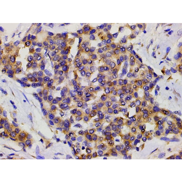
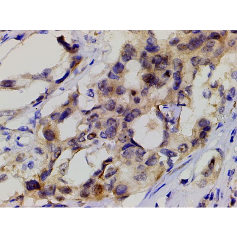
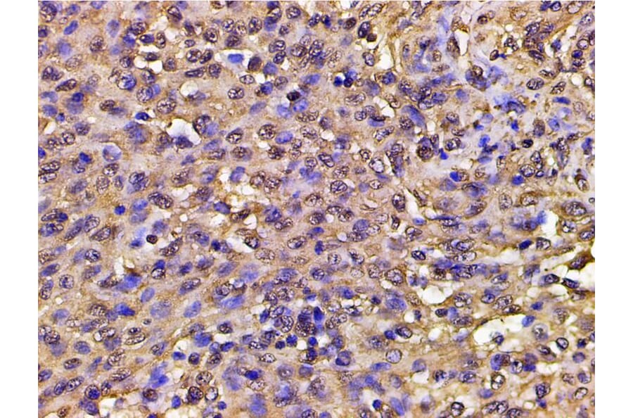
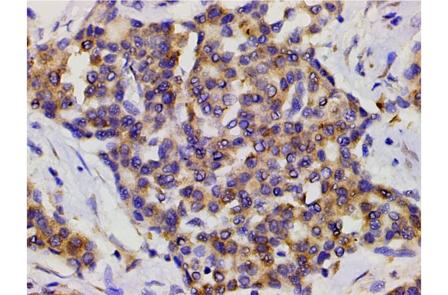
AF3241 at 1/200 staining human lung cancer tissue sections by IHC-P. The tissue was formaldehyde fixed and a heat mediated antigen retrieval step in citrate buffer was performed. The tissue was then blocked and incubated with the antibody for 1.5 hours at 22°C. An HRP conjugated goat anti-rabbit antibody was used as the secondary antibody.
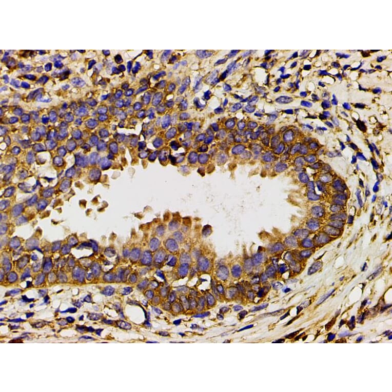
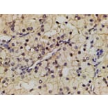
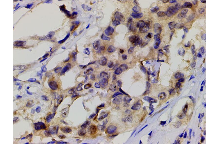
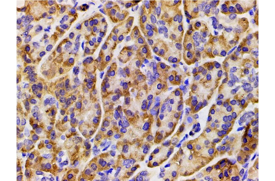
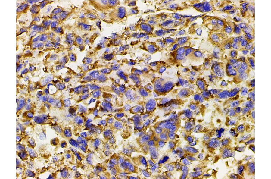
AF3241 at 1/200 staining human renal clear cell carcinoma tissue sections by IHC-P. The tissue was formaldehyde fixed and a heat mediated antigen retrieval step in citrate buffer was performed. The tissue was then blocked and incubated with the antibody for 1.5 hours at 22°C. An HRP conjugated goat anti-rabbit antibody was used as the secondary antibody.
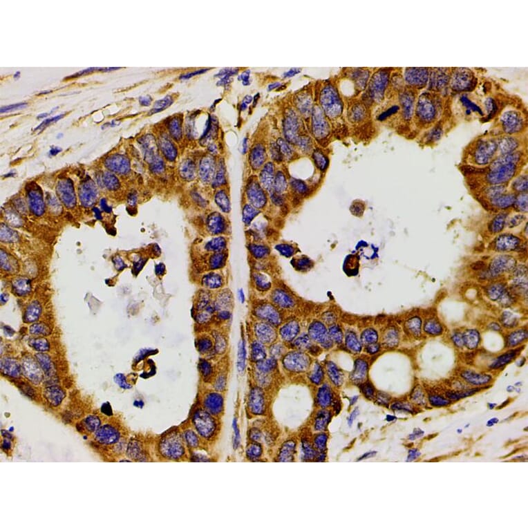
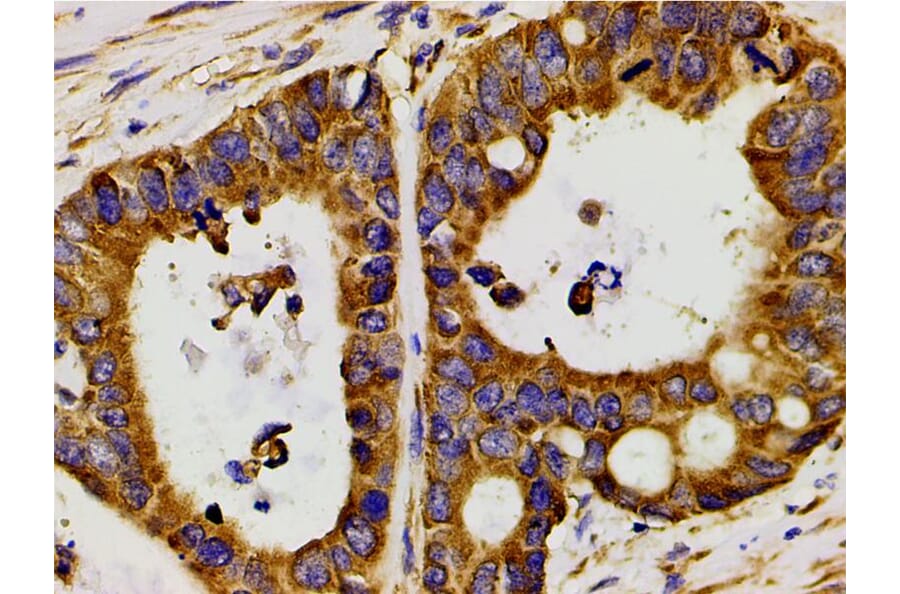
AF3241 at 1/200 staining human colon cancer tissue sections by IHC-P. The tissue was formaldehyde fixed and a heat mediated antigen retrieval step in citrate buffer was performed. The tissue was then blocked and incubated with the antibody for 1.5 hours at 22°C. An HRP conjugated goat anti-rabbit antibody was used as the secondary antibody.
AF3241 at 1/200 staining human duodenum tissue sections by IHC-P. The tissue was formaldehyde fixed and a heat mediated antigen retrieval step in citrate buffer was performed. The tissue was then blocked and incubated with the antibody for 1.5 hours at 22°C. An HRP conjugated goat anti-rabbit antibody was used as the secondary antibody.
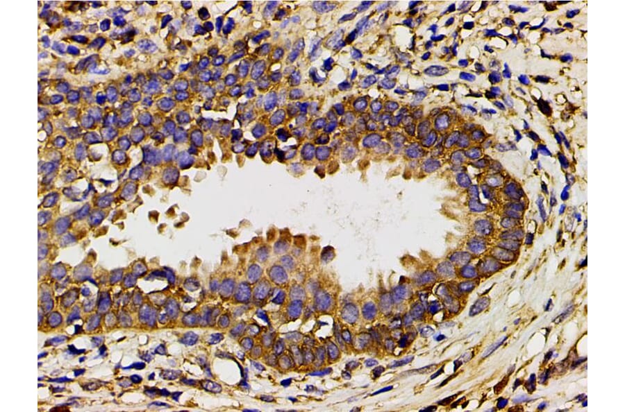
AF3241 at 1/200 staining human bladder cancer tissue sections by IHC-P. The tissue was formaldehyde fixed and a heat mediated antigen retrieval step in citrate buffer was performed. The tissue was then blocked and incubated with the antibody for 1.5 hours at 22°C. An HRP conjugated goat anti-rabbit antibody was used as the secondary antibody.
AF3241 at 1/200 staining human placenta tissue sections by IHC-P. The tissue was formaldehyde fixed and a heat mediated antigen retrieval step in citrate buffer was performed. The tissue was then blocked and incubated with the antibody for 1.5 hours at 22°C. An HRP conjugated goat anti-rabbit antibody was used as the secondary antibody.
AF3241 at 1/200 staining human vascular cancer tissue sections by IHC-P. The tissue was formaldehyde fixed and a heat mediated antigen retrieval step in citrate buffer was performed. The tissue was then blocked and incubated with the antibody for 1.5 hours at 22°C. An HRP conjugated goat anti-rabbit antibody was used as the secondary antibody.
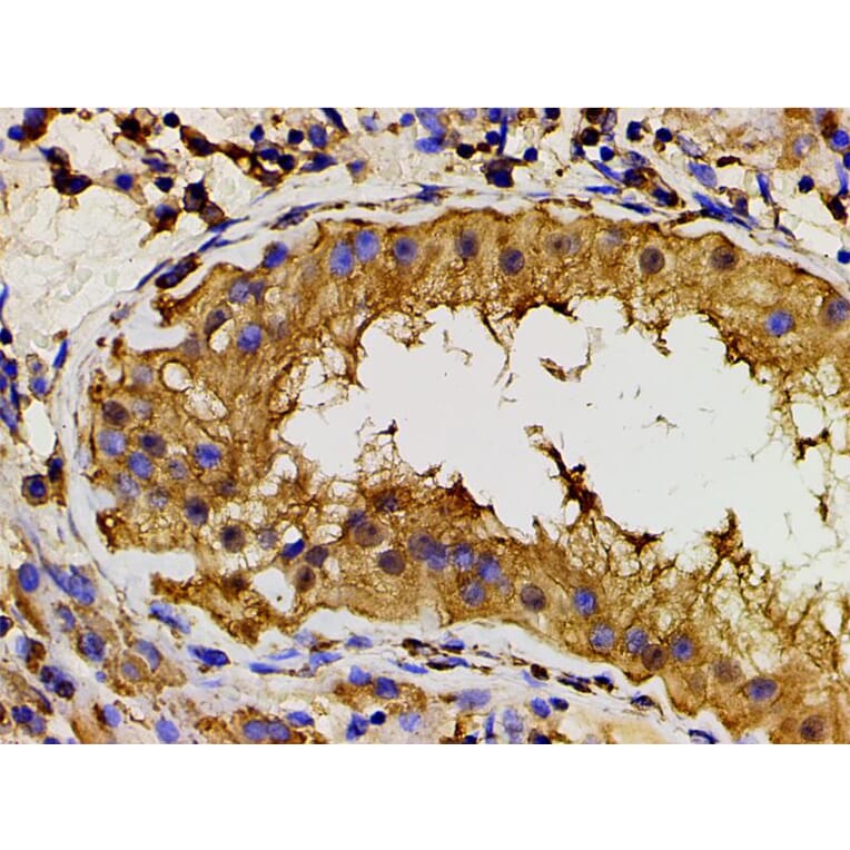
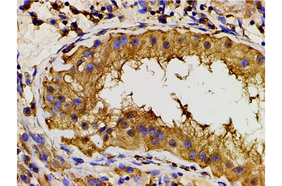
AF3241 at 1/200 staining human seminoma tissue sections by IHC-P. The tissue was formaldehyde fixed and a heat mediated antigen retrieval step in citrate buffer was performed. The tissue was then blocked and incubated with the antibody for 1.5 hours at 22°C. An HRP conjugated goat anti-rabbit antibody was used as the secondary antibody.
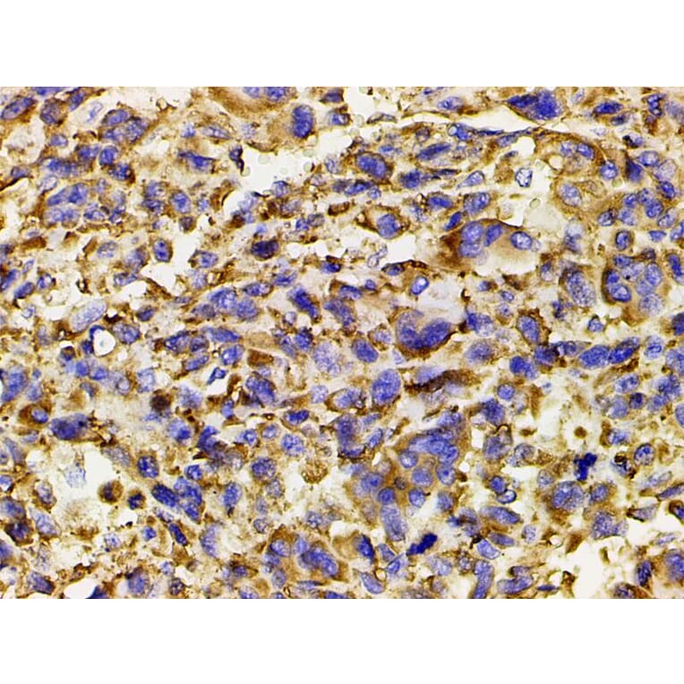
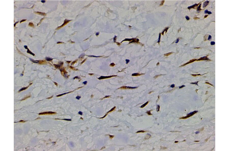
AF3241 at 1/200 staining human myosarcoma tissue sections by IHC-P. The tissue was formaldehyde fixed and a heat mediated antigen retrieval step in citrate buffer was performed. The tissue was then blocked and incubated with the antibody for 1.5 hours at 22°C. An HRP conjugated goat anti-rabbit antibody was used as the secondary antibody.
AF3241 at 1/200 staining human osteosarcoma tissue sections by IHC-P. The tissue was formaldehyde fixed and a heat mediated antigen retrieval step in citrate buffer was performed. The tissue was then blocked and incubated with the antibody for 1.5 hours at 22°C. An HRP conjugated goat anti-rabbit antibody was used as the secondary antibody.
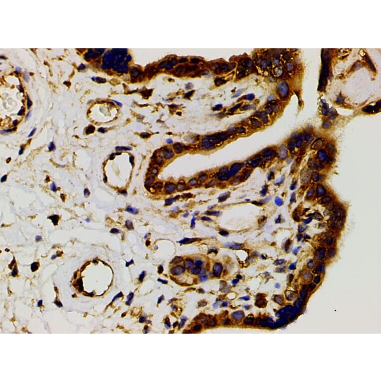
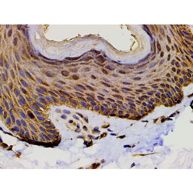
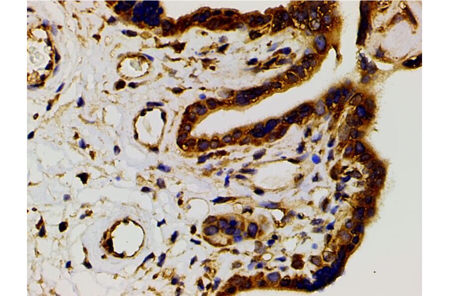
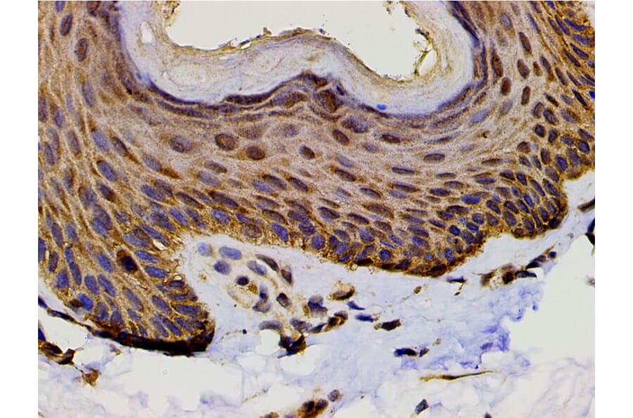
AF3241 at 1/200 staining human skin tissue sections by IHC-P. The tissue was formaldehyde fixed and a heat mediated antigen retrieval step in citrate buffer was performed. The tissue was then blocked and incubated with the antibody for 1.5 hours at 22°C. An HRP conjugated goat anti-rabbit antibody was used as the secondary antibody.
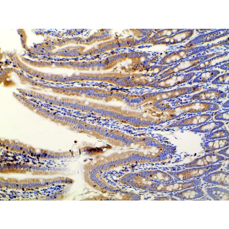
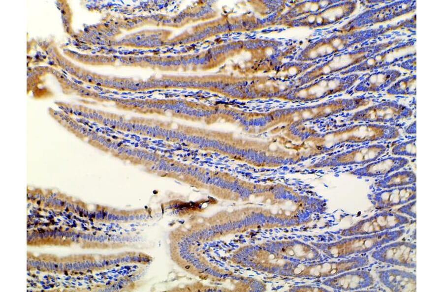
AF3241 at 1/100 staining rat Intestinal tissue sections by IHC-P. The tissue was formaldehyde fixed and a heat mediated antigen retrieval step in citrate buffer was performed. The tissue was then blocked and incubated with the antibody for 1.5 hours at 22°C. An HRP conjugated goat anti-rabbit antibody was used as the secondary antibody.
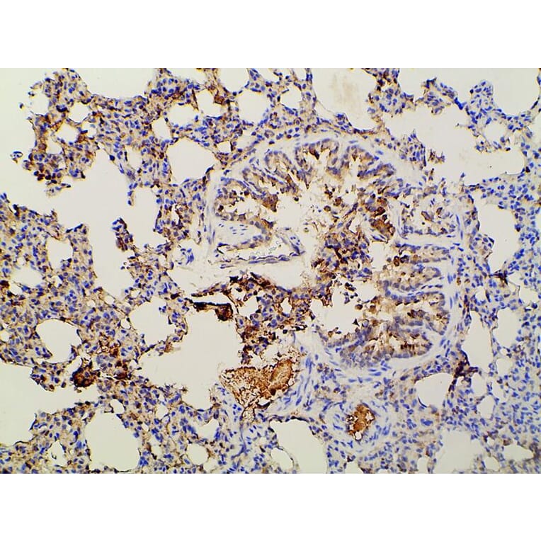
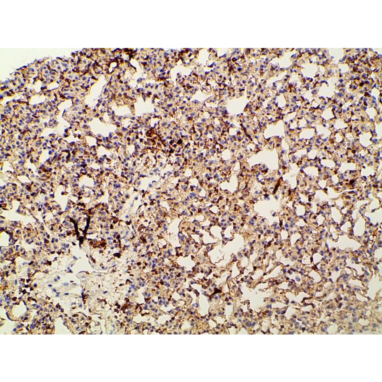
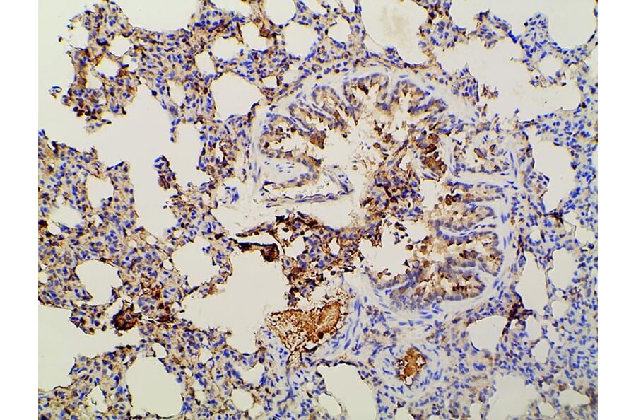
AF3241 at 1/100 staining rat lung tissue sections by IHC-P. The tissue was formaldehyde fixed and a heat mediated antigen retrieval step in citrate buffer was performed. The tissue was then blocked and incubated with the antibody for 1.5 hours at 22°C. An HRP conjugated goat anti-rabbit antibody was used as the secondary antibody.
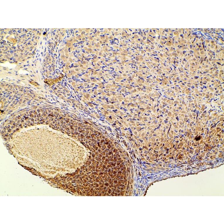
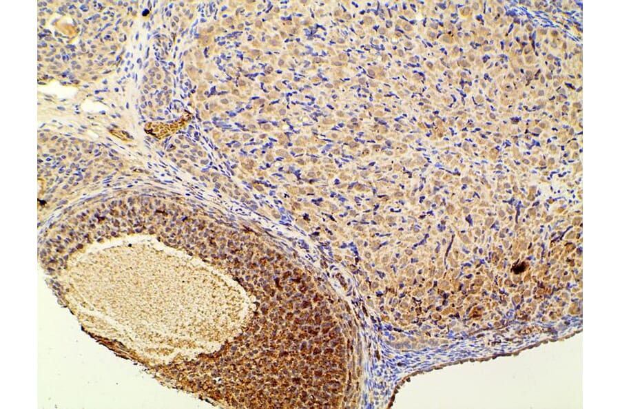
AF3241 at 1/100 staining rat ovarian tissue sections by IHC-P. The tissue was formaldehyde fixed and a heat mediated antigen retrieval step in citrate buffer was performed. The tissue was then blocked and incubated with the antibody for 1.5 hours at 22°C. An HRP conjugated goat anti-rabbit antibody was used as the secondary antibody.
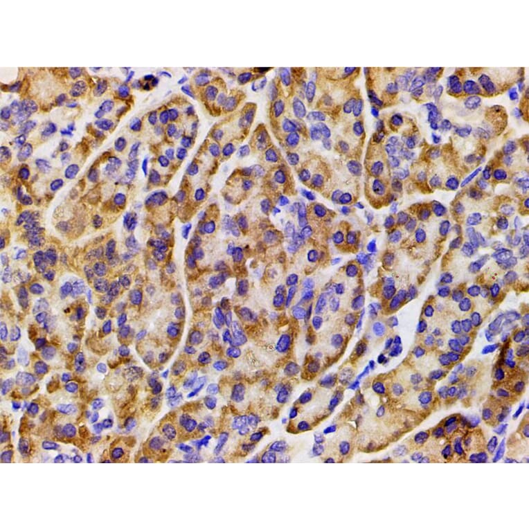
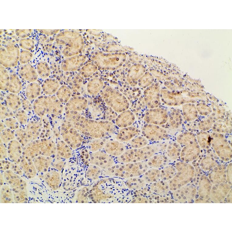
AF3241 at 1/100 staining rat kidney tissue sections by IHC-P. The tissue was formaldehyde fixed and a heat mediated antigen retrieval step in citrate buffer was performed. The tissue was then blocked and incubated with the antibody for 1.5 hours at 22°C. An HRP conjugated goat anti-rabbit antibody was used as the secondary antibody.
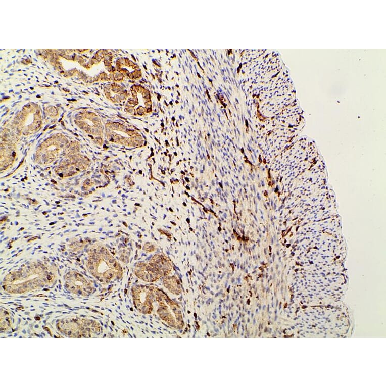
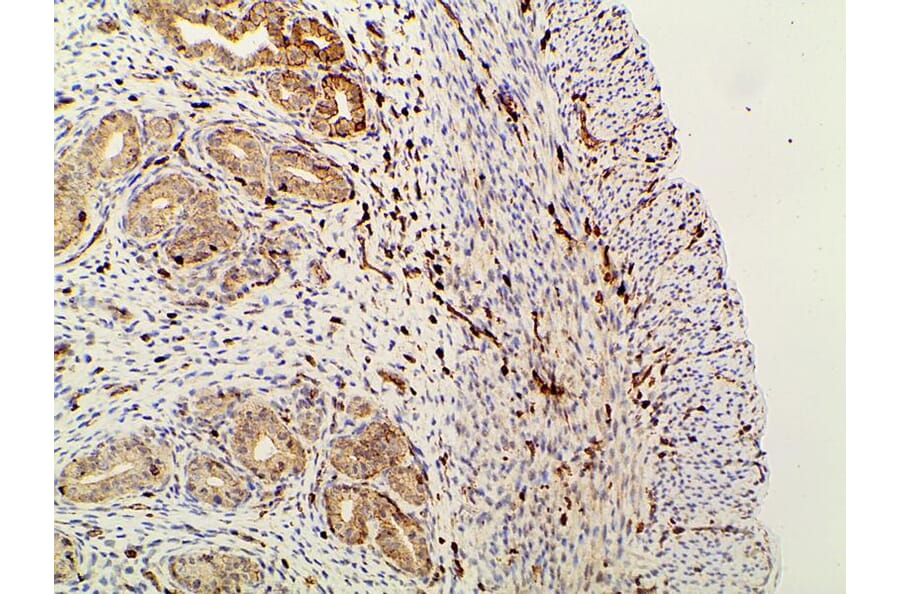
AF3241 at 1/100 staining rat gastric tissue sections by IHC-P. The tissue was formaldehyde fixed and a heat mediated antigen retrieval step in citrate buffer was performed. The tissue was then blocked and incubated with the antibody for 1.5 hours at 22°C. An HRP conjugated goat anti-rabbit antibody was used as the secondary antibody.
AF3241 at 1/100 staining rat uterine tissue sections by IHC-P. The tissue was formaldehyde fixed and a heat mediated antigen retrieval step in citrate buffer was performed. The tissue was then blocked and incubated with the antibody for 1.5 hours at 22°C. An HRP conjugated goat anti-rabbit antibody was used as the secondary antibody.
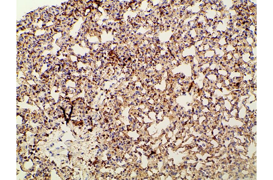
AF3241 at 1/100 staining mouse lung tissue sections by IHC-P. The tissue was formaldehyde fixed and a heat mediated antigen retrieval step in citrate buffer was performed. The tissue was then blocked and incubated with the antibody for 1.5 hours at 22°C. An HRP conjugated goat anti-rabbit antibody was used as the secondary antibody.
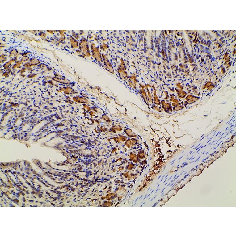
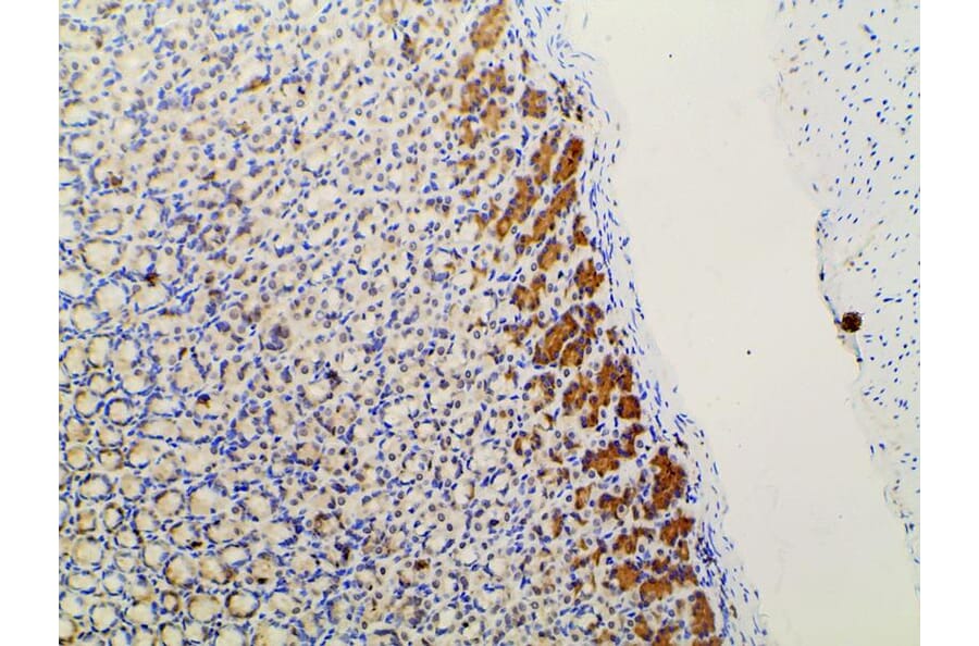
AF3241 at 1/100 staining mouse gastric tissue sections by IHC-P. The tissue was formaldehyde fixed and a heat mediated antigen retrieval step in citrate buffer was performed. The tissue was then blocked and incubated with the antibody for 1.5 hours at 22°C. An HRP conjugated goat anti-rabbit antibody was used as the secondary antibody.
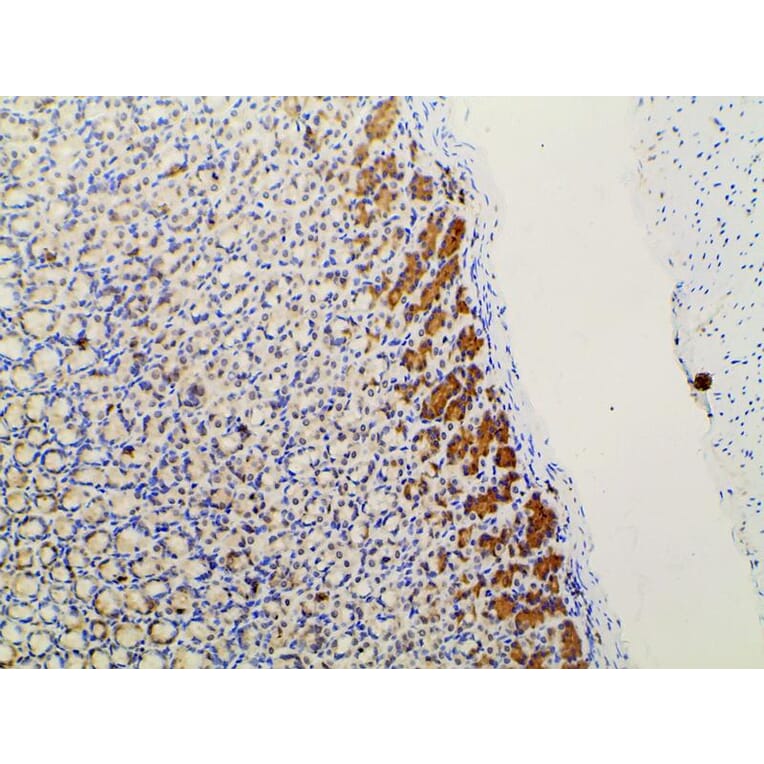
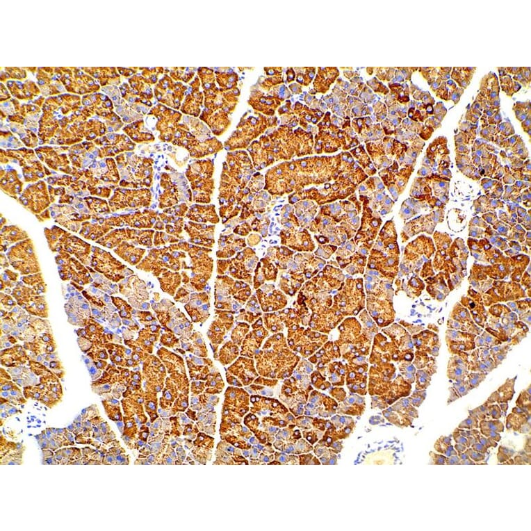
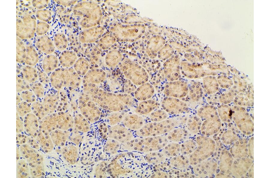
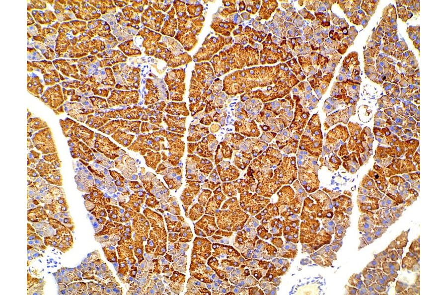
AF3241 at 1/100 staining mouse pancreatic tissue sections by IHC-P. The tissue was formaldehyde fixed and a heat mediated antigen retrieval step in citrate buffer was performed. The tissue was then blocked and incubated with the antibody for 1.5 hours at 22°C. An HRP conjugated goat anti-rabbit antibody was used as the secondary antibody.
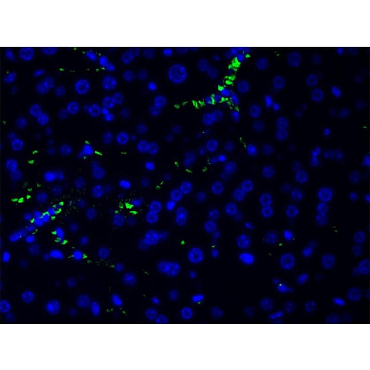
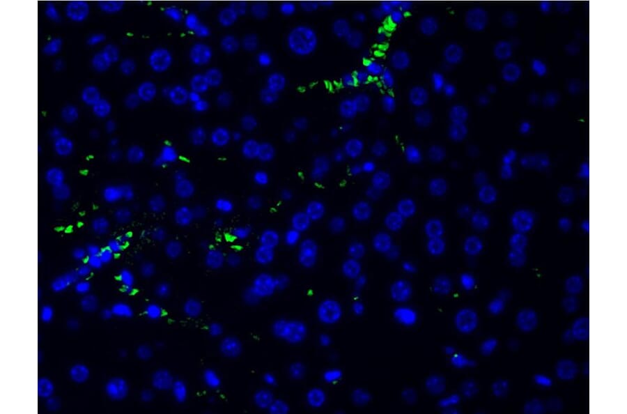
AF3241 at 1/200 staining human frozen liver tissue section by IHC-Fr. The sample was incubated with the primary antibody (1/200 in BSA) for 1 hour. An Alexa Fluor 488®-conjugated Goat anti-rabbit antibody was used as the secondary.
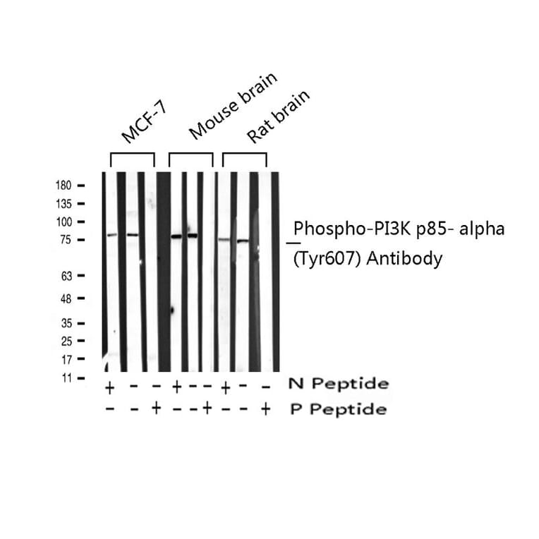
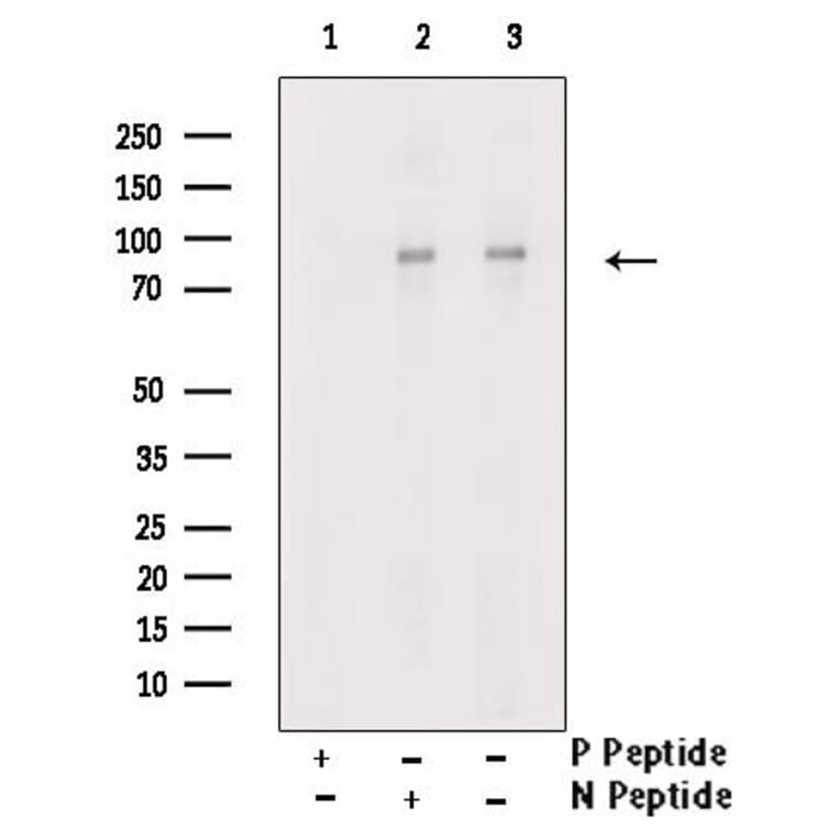
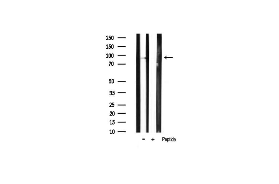
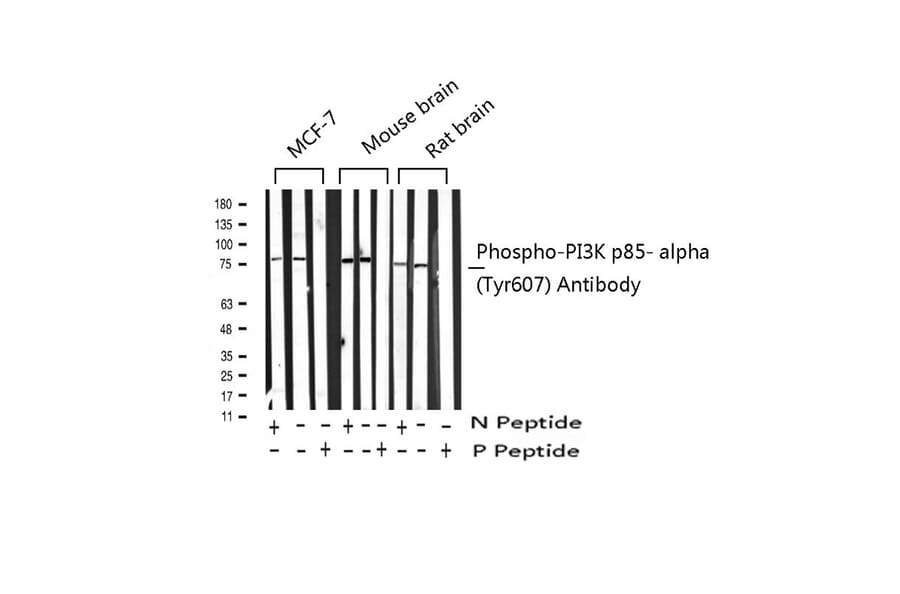
Western blot analysis of extracts from MCF7/mouse brain/rat brain, using Phospho-PI3K p85 alpha (Tyr607) Antibody. -/+ means absence or presence of N peptide(non-phospho peptide) and P peptide(phospho peptide)
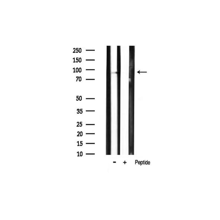
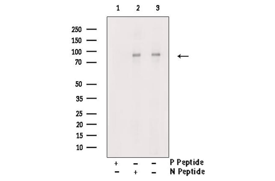
Western blot analysis of extracts from H2O2 treated Hela cells, using Phospho-PI3K p85 alpha (Tyr607) Antibody. Lane1 was treated with phospho-blocking peptide, Lane2 was treated with non-phospho-blocking peptide.
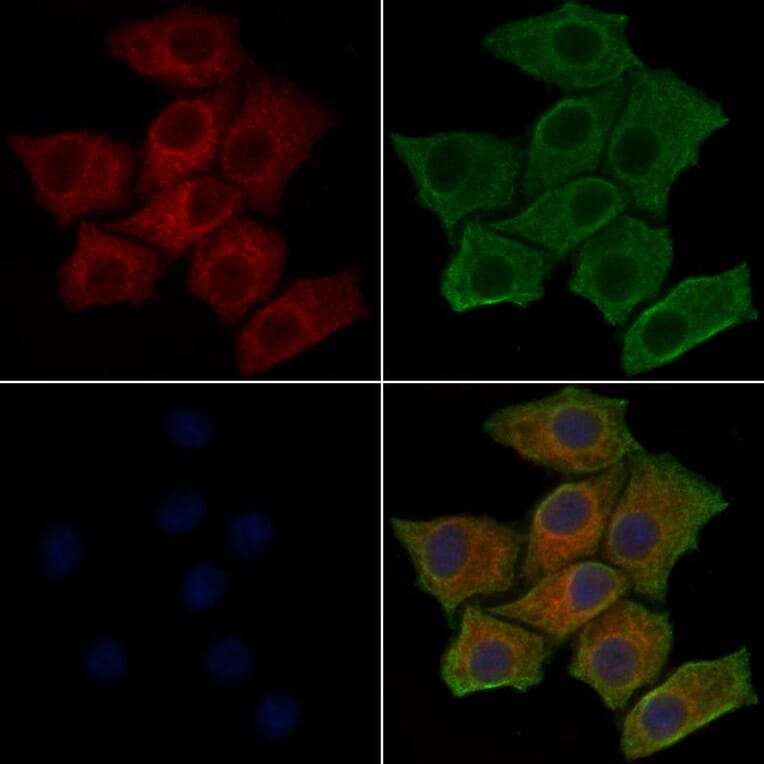
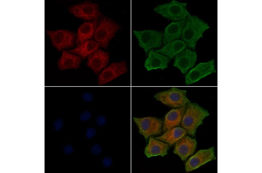
AF3241 staining HepG2 cells(30min of 4uM Forskolin treatment) by IF/ICC. The samples were fixed with PFA and permeabilized in 0.1% Triton X-100,then blocked in 10% serum for 45 minutes at 25°C. Samples were then incubated with primary Ab(AF3241) and mouse anti-beta tubulin Ab(T0023) for 1 hour at 37°C. An AlexaFluor594 conjugated goat anti-rabbit IgG(H+L) Ab(Red) and an AlexaFluor488 conjugated goat anti-mouse IgG(H+L) Ab(Green) were used as the secondary antibody. The nuclear counter stain is DAPI(blue).