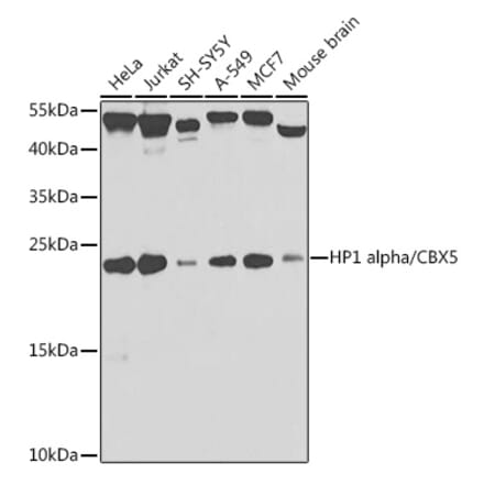
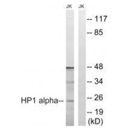
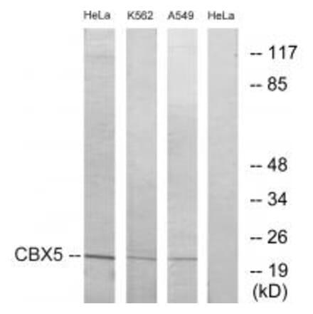
During the first half of the 20th century, biologist Emil Heitz used a new method of cytological staining to reveal that chromatin isn’t homogeneously distributed in the nucleus. Differences in the density of staining indicated that chromatin can be divided into two overarching categories based on their level of condensation: euchromatin and heterochromatin. We now understand that the spatial organization, condensation state and activity of chromatin within the nucleus are highly regulated and influence DNA replication, chromosome segregation, gene expression, DNA repair, and other fundamental biological activities. Unlike euchromatin, heterochromatic regions of the genome are highly condensed. The formation, spread and maintenance of heterochromatin are regulated by post-translational modifications (PTMs) and chromatin-modifying proteins. Based on the particular combination of PTMs and associated proteins, heterochromatin can be subdivided into two functional categories: facultative and constitutive. These two forms of heterochromatin are found in topologically distinct sub-compartments of the nucleus. Constitutive heterochromatin is typically found in association with centromeres and telomeres, which are gene-poor and contain repetitive DNA sequences. These regions are enriched with trimethylation of lysine 9 of histone H3 (H3K9me3), H3K20me3, heterochromatin protein 1 (HP1), and SUV4-20H, which together drive chromatin compaction. In contrast, facultative heterochromatin is marked by H3K27me3 and associated with Polycomb group (PcG) proteins. Facultative heterochromatin plays a crucial role in the silencing of developmentally-regulated genes. In eukaryotes, heterochromatin forms a unique domain at the nuclear periphery, where it is tethered at lamina-associated domains (LADs) by components of the nuclear lamina. In addition to modulating gene silencing during cell differentiation, heterochromatin is also known to serve a mechanical role in the nucleus and function in the DNA damage response. As a result, alterations in heterochromatin function are linked to human disease. We offer a range of heterochromatin markers including HP1 alpha antibodies and TRF1 antibodies, that are validated for use in multiple applications and available in various host species, antibody types, and formulations.
Anti-HP1 alpha Antibody (A82447) | |
100µg/$525 | |
| Description: | Goat polyclonal antibody to HP1 alpha. |
| Applications: | ELISA, WB, IF, Flow Cytometry |
| Reactivity: | Human |
| Conjugate: | Unconjugated |

Anti-HP1 alpha Antibody (A13174) | |
100µl/$375 | |
| Description: | Rabbit polyclonal antibody to HP1 alpha. |
| Applications: | WB, IHC-P, ICC/IF, IP, ChIP, ELISA |
| Reactivity: | Human, Mouse, Rat |
| Conjugate: | Unconjugated |
Anti-HP1 alpha Antibody [ARC0244] (A308742) | |
100µl/$515 | |
| Description: | Rabbit monoclonal [ARC0244] antibody to HP1 alpha. |
| Applications: | WB, IHC, ICC/IF, IP |
| Reactivity: | Human, Mouse, Rat |
| Conjugate: | Unconjugated |
Anti-HP1 alpha (phospho Ser92) Antibody (A95122) | |
10µg - 100µg/$200 – $505 | |
| Description: | Rabbit polyclonal antibody to HP1 alpha (phospho Ser92). |
| Applications: | IHC, IF, ELISA |
| Reactivity: | Human, Rat, Mouse |
| Conjugate: | Unconjugated |
Anti-HP1 alpha Antibody (A98569) | |
10µg - 100µg/$200 – $505 | |
| Description: | Rabbit polyclonal antibody to HP1 alpha. |
| Applications: | WB,IHC-p,IF(paraffin section),ELISA |
| Reactivity: | Human,Rat,Mouse |
| Conjugate: | Unconjugated |
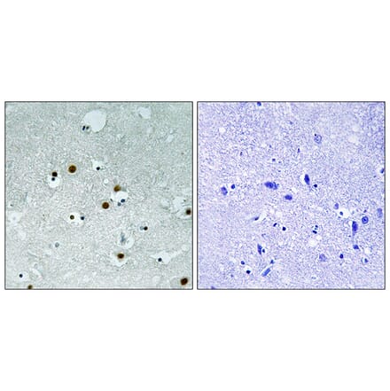
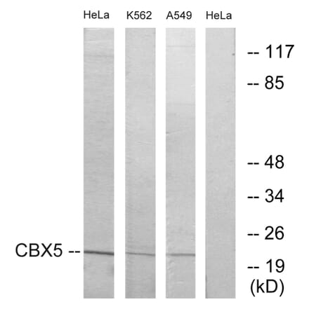
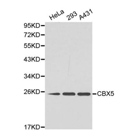
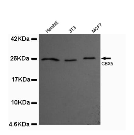
Anti-CBX5 Antibody (A99464) | |
10µg - 100µg/$200 – $505 | |
| Description: | Rabbit polyclonal antibody to CBX5. |
| Applications: | WB, IHC, IF, ELISA |
| Reactivity: | Human, Mouse |
| Conjugate: | Unconjugated |
Anti-CBX5 (K69) Antibody (A25975) | |
50µl - 100µl/$415 – $560 | |
| Description: | Rabbit polyclonal antibody to CBX5 (K69) |
| Applications: | WB, IHC, IF |
| Reactivity: | Human, Mouse, Rat |
| Conjugate: | Unconjugated |
Anti-CBX5 Antibody (A34214) | |
50µl - 100µl/$415 – $595 | |
| Description: | Rabbit polyclonal antibody to CBX5 |
| Applications: | WB, ICC/IF |
| Reactivity: | Human, Rat |
| Conjugate: | Unconjugated |
Anti-CBX5 Antibody (A40176) | |
100µl/$430 | |
| Description: | Mouse monoclonal antibody to CBX5 |
| Applications: | WB, ICC/IF |
| Reactivity: | Human, Mouse, Rat, Canine |
| Conjugate: | Unconjugated |
Anti-HP1 alpha Antibody (A88778) | |
100µl/$5151 Citation | |
| Description: | Rabbit polyclonal antibody to HP1 alpha. |
| Applications: | WB, ICC/IF |
| Reactivity: | Human, Mouse |
| Conjugate: | Unconjugated |
Anti-HP1 alpha Antibody [24GB3210] (A346549) | |
100µl/$450 | |
| Description: | Recombinant rabbit monoclonal [24GB3210] antibody to HP1 alpha. |
| Applications: | WB, Flow Cytometry, ICC/IF |
| Reactivity: | Human, Mouse, Rat |
| Conjugate: | Unconjugated |
Anti-HP1 alpha Antibody [25GB2865] (A346550) | |
100µl/$640 | |
| Description: | Recombinant rabbit monoclonal [25GB2865] antibody to HP1 alpha. |
| Applications: | WB, Flow Cytometry, ICC/IF |
| Reactivity: | Human, Mouse, Rat |
| Conjugate: | Unconjugated |
Anti-HP1 alpha (Ab-92) Antibody (A36281) | |
50µl - 100µl/$290 – $430 | |
| Description: | Rabbit polyclonal antibody to HP1 alpha (Ab-92) |
| Applications: | WB, IHC |
| Reactivity: | Human |
| Conjugate: | Unconjugated |
Anti-HP1 alpha Antibody (A372) | |
50µg/$405 | |
| Description: | Rabbit polyclonal antibody to HP1 alpha. |
| Applications: | WB, IF |
| Reactivity: | Human |
| Conjugate: | Unconjugated |
Anti-CBX5 Antibody (A43649) | |
50µl - 100µl/$290 – $430 | |
| Description: | Rabbit polyclonal antibody to CBX5 |
| Applications: | WB |
| Reactivity: | Human, Mouse |
| Conjugate: | Unconjugated |
Anti-CBX5 Antibody (A49750) | |
100µl/$430 | |
| Description: | Rabbit polyclonal antibody to CBX5 |
| Applications: | ELISA, WB |
| Reactivity: | Human |
| Conjugate: | Unconjugated |
Anti-HP1 alpha Antibody (Biotin) (A83586) | |
100µg/$525 | |
| Description: | Goat polyclonal antibody to HP1 alpha (Biotin). |
| Applications: | ELISA, WB |
| Reactivity: | Human |
| Conjugate: | Biotin |
Showing 1-17 of 17 products