Supplied in Phosphate Buffered Saline, pH 7.3, with 50% Glycerol, 0.05% BSA, and 0.05% Proclin 300.
Storage
Shipped at 4°C. Upon delivery aliquot and store at -20°C. Avoid freeze / thaw cycles.
Synonyms
GTP binding protein RAB10, RAB10 member RAS oncogene family, RAB10_HUMAN, Ras related GTP binding protein, Ras related GTP binding protein RAB10, Ras-related protein Rab-10
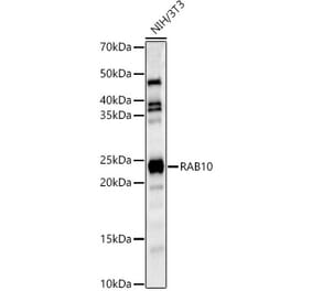
Figure 1: Western Blot - Anti-RAB10 Antibody [ARC55653] (A309454)
Western blot analysis of various lysates, using Anti-RAB10 Antibody [ARC55653] (A309454) at 1:60,000 dilution. The secondary antibody was Goat Anti-Rabbit IgG H&L Antibody (HRP) at 1:10,000 dilution. Lysates/proteins were present at 25µg per lane. The blocking buffer used was 3% non-fat dry milk in TBST. Detection was with a ECL Basic Kit. Exposure time: 180s.
Figure 2: Western Blot - Anti-RAB10 Antibody [ARC55653] (A309454)
Western blot analysis of extracts from wild type(WT) and RAB10 knockdown (KD) U-87MG(KD) cells, using Anti-RAB10 Antibody [ARC55653] (A309454) at 1:60,000 dilution. The secondary antibody was Goat Anti-Rabbit IgG H&L Antibody (HRP) at 1:10,000 dilution. Lysates/proteins were present at 25µg per lane. The blocking buffer used was 3% non-fat dry milk in TBST. Detection was with a ECL Basic Kit. Exposure time: 180s.
Immunofluorescence analysis of MCF7 using Anti-RAB10 Antibody [ARC55653] (A309454) at a dilution of 1:200 (40x lens). DAPI was used to stain the cell nuclei (blue).
Immunofluorescence analysis of NIH/3T3 using Anti-RAB10 Antibody [ARC55653] (A309454) at a dilution of 1:200 (40x lens). DAPI was used to stain the cell nuclei (blue).
Publishing research using Anti-RAB10 Antibody [ARC55653] (A309454)? Please let us know so that we can list the citation on this page.
Alternative products to Anti-RAB10 Antibody [ARC55653] (A309454)


![Western Blot - Anti-RAB10 Antibody [ARC55653] (A309454) - Antibodies.com](https://cdn.antibodies.com/image/catalog/309/A309454_1.jpg?profile=product_top)
![Western Blot - Anti-RAB10 Antibody [ARC55653] (A309454) - Antibodies.com](https://cdn.antibodies.com/image/catalog/309/A309454_2.jpg?profile=product_top)
![Immunofluorescence - Anti-RAB10 Antibody [ARC55653] (A309454) - Antibodies.com](https://cdn.antibodies.com/image/catalog/309/A309454_3.jpg?profile=product_top)
![Immunofluorescence - Anti-RAB10 Antibody [ARC55653] (A309454) - Antibodies.com](https://cdn.antibodies.com/image/catalog/309/A309454_4.jpg?profile=product_top)
![Western Blot - Anti-RAB10 Antibody [ARC55653] (A309454) - Antibodies.com](https://cdn.antibodies.com/image/catalog/309/A309454_1.jpg?profile=product_top_thumb)
![Western Blot - Anti-RAB10 Antibody [ARC55653] (A309454) - Antibodies.com](https://cdn.antibodies.com/image/catalog/309/A309454_2.jpg?profile=product_top_thumb)
![Immunofluorescence - Anti-RAB10 Antibody [ARC55653] (A309454) - Antibodies.com](https://cdn.antibodies.com/image/catalog/309/A309454_3.jpg?profile=product_top_thumb)
![Immunofluorescence - Anti-RAB10 Antibody [ARC55653] (A309454) - Antibodies.com](https://cdn.antibodies.com/image/catalog/309/A309454_4.jpg?profile=product_top_thumb)
![Western Blot - Anti-RAB10 Antibody [ARC55653] (A309454) - Antibodies.com](https://cdn.antibodies.com/image/catalog/309/A309454_1.jpg?profile=product_image)
![Western Blot - Anti-RAB10 Antibody [ARC55653] (A309454) - Antibodies.com](https://cdn.antibodies.com/image/catalog/309/A309454_2.jpg?profile=product_image)
![Immunofluorescence - Anti-RAB10 Antibody [ARC55653] (A309454) - Antibodies.com](https://cdn.antibodies.com/image/catalog/309/A309454_3.jpg?profile=product_image)
![Immunofluorescence - Anti-RAB10 Antibody [ARC55653] (A309454) - Antibodies.com](https://cdn.antibodies.com/image/catalog/309/A309454_4.jpg?profile=product_image)