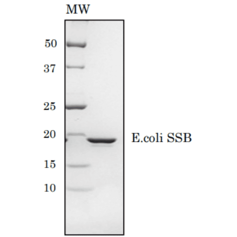
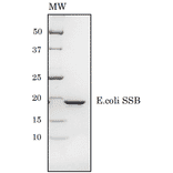
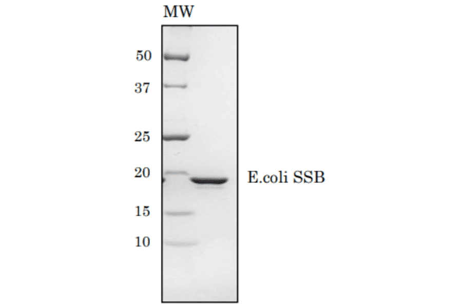
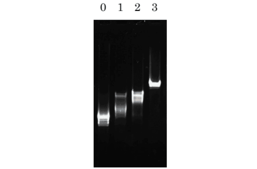
Functional single-stranded DNA-binding protein for studying DNA replication and recombination.
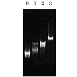
Enhancement of the specificity and yield of PCR.
The complex formation between the single-strand DNA binding protein (ssB protein) from Escherichia coli and oligonucleotides and single-stranded DNA has been studied by using fluorescence titrations, ultracentrifugation measurements, and fast kinetic techniques. Determination of the stoichiometries of oligo(dT)--ssB complexes shows that each of the four subunits of the ssB protein represents a binding site for an oligonucleotide about eight residues long. Occupation of all four binding sites with oligo(dT) or poly(dT) leads to 80% quenching of the intrinsic protein fluorescence. The binding sites are nearly equivalent and independent. For d(pT)16, the intrinsic binding constant is 6 X 10(5) M-1, and for d(pT)30-40, which is long enough to extend continuously over the ssB tetramer, the binding constant is higher than 5 X 10(8) M-1. Oligoadenylates bind about 2 orders of magnitude weaker than the corresponding oligo(dT) species. The binding of oligo(dT) is very weakly dependent on ionic strength, in contrast to the oligo(dA)--ssB complex formation. For d(pT)8, d(pT)16, and d(pT)30-40, the complex formation can be described by a simple one-step reaction. The strength of the interaction is mainly expressed in the rate constant of dissociation. In the cooperative complexes with poly(dT) or poly(dA), all four binding sites on the ssB tetramer are also occupied. It is concluded that single-stranded DNA is coiled around the ssB molecule. Fluorescence melting experiments of the complexes show that the conformation of the single-stranded DNA has a strong influence on the stability of the complexes.
The DNA unwinding protein of Escherichia coli (Sigal, N., Delius, H., Kornberg, T., Gefter, M., and Alberts, B. (1972) Proc. Nat. Acad. Sci. U.S.A. 69, 3537-3541) has been purified to homogeneity by a simple procedure which utilizes its stability to heating. The protein is an asymmetric tetramer of 18,500 dalton subunits which binds preferentially to single-stranded DNA at a ratio of one protein molecule per 32 nucleotides. Binding to DNA is complete in less than 10 s at 0 degrees while release of the protein from single-stranded DNA is relatively slow even at 37 degrees. A simple functional assay for unwinding protein depends on its essential role in the conversion of phage G4 single-stranded DNA to the replicative form. Unwinding protein stimulates initiation of replication of all single-stranded phage DNAs. Approximately 300 copies of unwinding protein are present per cell, as estimated by antibody titration, an amount sufficient to cover substantial lengths of DNA in several replicating forks.