Supplied in Phosphate Buffered Saline, pH 7.3, with 50% Glycerol, 0.05% BSA, and 0.05% Proclin 300.
Storage
Shipped at 4°C. Upon delivery aliquot and store at -20°C. Avoid freeze / thaw cycles.
Synonyms
DNA binding protein SATB2, DNA-binding protein SATB2, FLJ21474, FLJ32076, GLSS, KIAA1034, MGC119474, MGC119477, SATB family member 2, SATB homeobox 2, SATB2_HUMAN, Special AT rich sequence binding protein 2, Special AT-rich sequence-binding protein 2
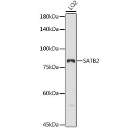
Figure 1: Western Blot - Anti-SATB2 Antibody [ARC50977] (A306174)
Western blot analysis of extracts of various cell lines, using Anti-SATB2 Antibody [ARC50977] (A306174) at 1:10,000 dilution. The secondary antibody was Goat Anti-Rabbit IgG H&L Antibody (HRP) at 1:10,000 dilution. Lysates/proteins were present at 25µg per lane. The blocking buffer used was 3% non-fat dry milk in TBST. Detection was with a ECL Basic Kit. Exposure time: 90s.
Immunohistochemistry analysis of paraffin-embedded human appendix tissue using Anti-SATB2 Antibody [ARC50977] (A306174) at a dilution of 1:2000(40x lens). Perform high pressure antigen retrieval with 10 mM citrate buffer pH 6.0 before commencing with IHC staining protocol.
Immunohistochemistry analysis of paraffin-embedded human pancreas (negative control sample) using Anti-SATB2 Antibody [ARC50977] (A306174) at a dilution of 1:2000(40x lens). Perform high pressure antigen retrieval with 10 mM citrate buffer pH 6.0 before commencing with IHC staining protocol.
Immunohistochemistry analysis of paraffin-embedded human colon carcinoma tissue using Anti-SATB2 Antibody [ARC50977] (A306174) at a dilution of 1:2000(40x lens). Perform high pressure antigen retrieval with 10 mM citrate buffer pH 6.0 before commencing with IHC staining protocol.
Immunofluorescence analysis of mouse brain using Anti-SATB2 Antibody [ARC50977] (A306174) at a dilution of 1:200 (40x lens). DAPI was used to stain the cell nuclei (blue).


![Western Blot - Anti-SATB2 Antibody [ARC50977] (A306174) - Antibodies.com](https://cdn.antibodies.com/image/catalog/306/A306174_1.jpg?profile=product_top)
![Immunohistochemistry - Anti-SATB2 Antibody [ARC50977] (A306174) - Antibodies.com](https://cdn.antibodies.com/image/catalog/306/A306174_2.jpg?profile=product_top)
![Immunohistochemistry - Anti-SATB2 Antibody [ARC50977] (A306174) - Antibodies.com](https://cdn.antibodies.com/image/catalog/306/A306174_3.jpg?profile=product_top)
![Immunohistochemistry - Anti-SATB2 Antibody [ARC50977] (A306174) - Antibodies.com](https://cdn.antibodies.com/image/catalog/306/A306174_4.jpg?profile=product_top)
![Immunofluorescence - Anti-SATB2 Antibody [ARC50977] (A306174) - Antibodies.com](https://cdn.antibodies.com/image/catalog/306/A306174_5.jpg?profile=product_top)
![Western Blot - Anti-SATB2 Antibody [ARC50977] (A306174) - Antibodies.com](https://cdn.antibodies.com/image/catalog/306/A306174_1.jpg?profile=product_top_thumb)
![Immunohistochemistry - Anti-SATB2 Antibody [ARC50977] (A306174) - Antibodies.com](https://cdn.antibodies.com/image/catalog/306/A306174_2.jpg?profile=product_top_thumb)
![Immunohistochemistry - Anti-SATB2 Antibody [ARC50977] (A306174) - Antibodies.com](https://cdn.antibodies.com/image/catalog/306/A306174_3.jpg?profile=product_top_thumb)
![Immunohistochemistry - Anti-SATB2 Antibody [ARC50977] (A306174) - Antibodies.com](https://cdn.antibodies.com/image/catalog/306/A306174_4.jpg?profile=product_top_thumb)
![Immunofluorescence - Anti-SATB2 Antibody [ARC50977] (A306174) - Antibodies.com](https://cdn.antibodies.com/image/catalog/306/A306174_5.jpg?profile=product_top_thumb)
![Western Blot - Anti-SATB2 Antibody [ARC50977] (A306174) - Antibodies.com](https://cdn.antibodies.com/image/catalog/306/A306174_1.jpg?profile=product_image)
![Immunohistochemistry - Anti-SATB2 Antibody [ARC50977] (A306174) - Antibodies.com](https://cdn.antibodies.com/image/catalog/306/A306174_2.jpg?profile=product_image)
![Immunohistochemistry - Anti-SATB2 Antibody [ARC50977] (A306174) - Antibodies.com](https://cdn.antibodies.com/image/catalog/306/A306174_3.jpg?profile=product_image)
![Immunohistochemistry - Anti-SATB2 Antibody [ARC50977] (A306174) - Antibodies.com](https://cdn.antibodies.com/image/catalog/306/A306174_4.jpg?profile=product_image)
![Immunofluorescence - Anti-SATB2 Antibody [ARC50977] (A306174) - Antibodies.com](https://cdn.antibodies.com/image/catalog/306/A306174_5.jpg?profile=product_image)
![Western Blot - Anti-SATB2 Antibody [ARC2363] (A308738) - Antibodies.com](https://cdn.antibodies.com/image/catalog/308/A308738_1.jpg?profile=product_alternative)
![Immunohistochemistry - Anti-SATB2 Antibody [RM365] (A121296) - Antibodies.com](https://cdn.antibodies.com/image/catalog/121/A121460_1.png?profile=product_alternative)
![Immunohistochemistry - Anti-SATB2 Antibody [rSATB2/6929] (A277985) - Antibodies.com](https://cdn.antibodies.com/image/catalog/277/A277985_1.jpg?profile=product_alternative)
![Immunohistochemistry - Anti-SATB2 Antibody [rSATB2/6929] - BSA and Azide free (A278573) - Antibodies.com](https://cdn.antibodies.com/image/catalog/278/A278573_1.jpg?profile=product_alternative)
![Immunohistochemistry - Anti-SATB2 Antibody [SATB2/4374R] - BSA and Azide free (A251760) - Antibodies.com](https://cdn.antibodies.com/image/catalog/251/A251760_1.jpg?profile=product_alternative)
![Immunohistochemistry - Anti-SATB2 Antibody [SATB2/4374R] (A248578) - Antibodies.com](https://cdn.antibodies.com/image/catalog/248/A248578_1.jpg?profile=product_alternative)

![Immunohistochemistry - Anti-SATB2 Antibody [IHC095] (A324610) - Antibodies.com](https://cdn.antibodies.com/image/catalog/324/A324610_1.png?profile=product_alternative)