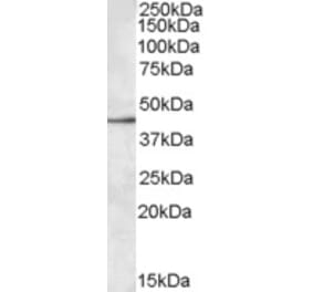
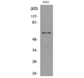
Figure 1: Western Blot - Anti-SIGLEC8 Antibody [ARC52425] (A307227)
Western blot analysis of extracts of normal 293T cells and 293T transfected with Siglec-8 protein, using Anti-SIGLEC8 Antibody [ARC52425] (A307227) at 1:2,000 dilution. The secondary antibody was Goat Anti-Rabbit IgG H&L Antibody (HRP) at 1:10,000 dilution. Lysates/proteins were present at 25µg per lane. The blocking buffer used was 3% non-fat dry milk in TBST. Detection was with a ECL Basic Kit. Exposure time: 90s.
Flow cytometry analysis of 293F cells transfected with Siglec8, stained with Rabbit IgG isotype control (10 µg/ml, blue line) or Anti-SIGLEC8 Antibody [ARC52425] (A307227), (10 µg/ml green line), followed by Alexa Fluor 647 conjugated goat anti-rabbit polyclonal antibody (1:600 dilution) staining. Non-fluorescently stained 293F cells transfected with Siglec8 cells, used as blank control (red line).
Flow cytometry analysis of 293F cells, stained with Rabbit IgG isotype control (10 µg/ml, blue line) or Anti-SIGLEC8 Antibody [ARC52425] (A307227), (10 µg/ml green line), followed by Alexa Fluor 647 conjugated goat anti-rabbit polyclonal antibody (1:600 dilution). Non-fluorescently stained 293F cells, used as blank control (red line).
Publishing research using Anti-SIGLEC8 Antibody [ARC52425] (A307227)? Please let us know so that we can list the citation on this page.
Alternative products to Anti-SIGLEC8 Antibody [ARC52425] (A307227)


![Western Blot - Anti-SIGLEC8 Antibody [ARC52425] (A307227) - Antibodies.com](https://cdn.antibodies.com/image/catalog/307/A307227_1.jpg?profile=product_top)
![Flow Cytometry - Anti-SIGLEC8 Antibody [ARC52425] (A307227) - Antibodies.com](https://cdn.antibodies.com/image/catalog/307/A307227_2.jpg?profile=product_top)
![Flow Cytometry - Anti-SIGLEC8 Antibody [ARC52425] (A307227) - Antibodies.com](https://cdn.antibodies.com/image/catalog/307/A307227_3.jpg?profile=product_top)
![Western Blot - Anti-SIGLEC8 Antibody [ARC52425] (A307227) - Antibodies.com](https://cdn.antibodies.com/image/catalog/307/A307227_1.jpg?profile=product_top_thumb)
![Flow Cytometry - Anti-SIGLEC8 Antibody [ARC52425] (A307227) - Antibodies.com](https://cdn.antibodies.com/image/catalog/307/A307227_2.jpg?profile=product_top_thumb)
![Flow Cytometry - Anti-SIGLEC8 Antibody [ARC52425] (A307227) - Antibodies.com](https://cdn.antibodies.com/image/catalog/307/A307227_3.jpg?profile=product_top_thumb)
![Western Blot - Anti-SIGLEC8 Antibody [ARC52425] (A307227) - Antibodies.com](https://cdn.antibodies.com/image/catalog/307/A307227_1.jpg?profile=product_image)
![Flow Cytometry - Anti-SIGLEC8 Antibody [ARC52425] (A307227) - Antibodies.com](https://cdn.antibodies.com/image/catalog/307/A307227_2.jpg?profile=product_image)
![Flow Cytometry - Anti-SIGLEC8 Antibody [ARC52425] (A307227) - Antibodies.com](https://cdn.antibodies.com/image/catalog/307/A307227_3.jpg?profile=product_image)
![Flow Cytometry - Anti-SIGLEC8 Antibody [7C9] (A242894) - Antibodies.com](https://cdn.antibodies.com/image/catalog/242/A242895_1.jpg?profile=product_alternative)