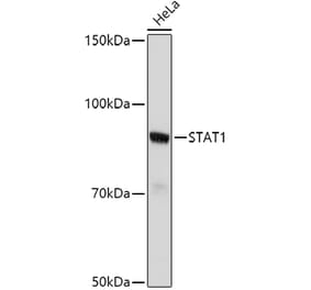
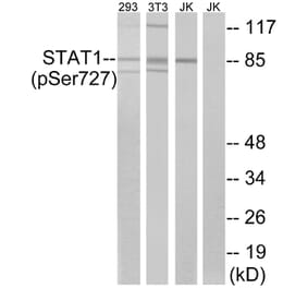
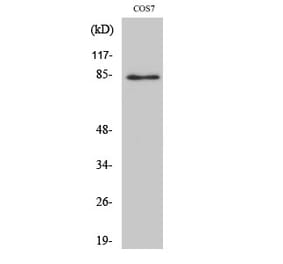
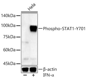
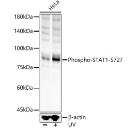
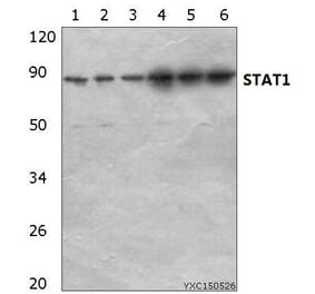
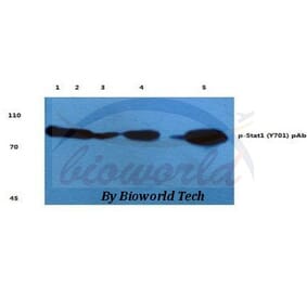
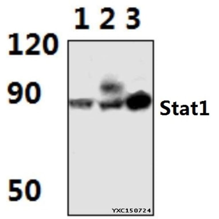
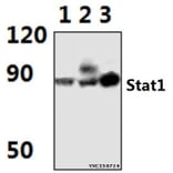
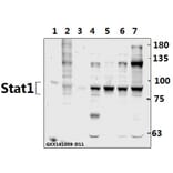
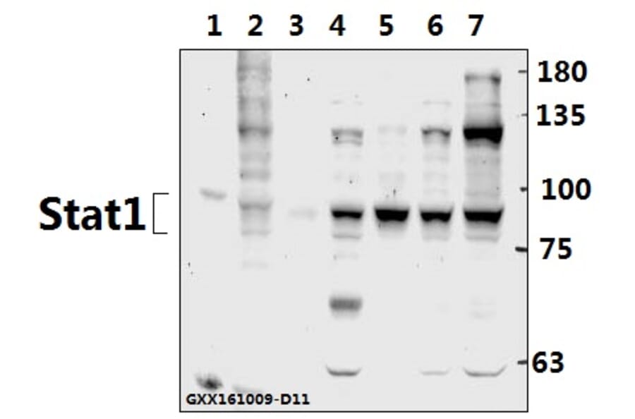
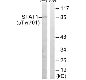
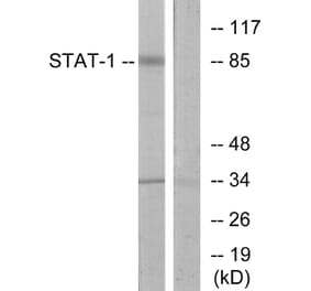
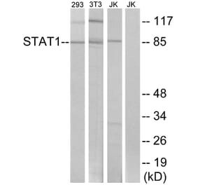
Stat1 (G695) pAb detects endogenous levels of Stat1 protein.
Applications
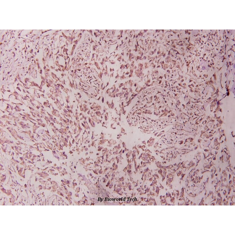
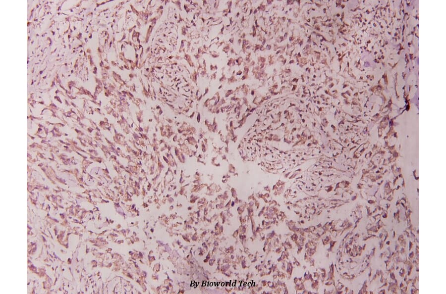
WB, IHC, IP
Reactivity
Human, Mouse, Rat
Immunogen
Synthetic peptide, corresponding to amino acids 680-720 of Human Stat1.
Host
Rabbit
Clonality
Polyclonal
Conjugate
Unconjugated
Molecular Weight
~ 87 kDa
Purity
The antibody was affinity-purified from rabbit antiserum by affinity-chromatography using epitope-specific immunogen and the purity is > 95% (by SDS-PAGE).
Product Form
1 mg/ml in Phosphate buffered saline (PBS) with 0.05% sodium azide, approx. pH 7.2.
Synonyms
Signal transducer and activator of transcription 1-alpha/beta, Transcription factor ISGF-3 components p91/p84


![WB - Anti-STAT1 Antibody [SM1] (A85870)](https://cdn.antibodies.com/image/catalog/85/A85870_1.jpg?profile=product_alternative)