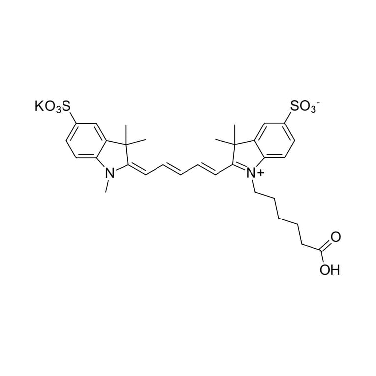
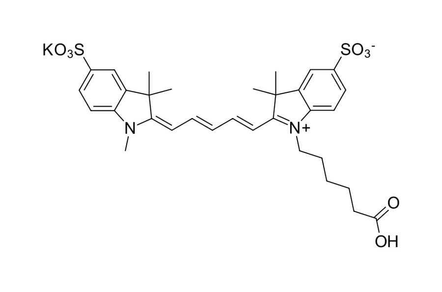
Non-activated sulfo-Cyanine 5 carboxylic acid, water soluble dye. This dye is highly hydrophilic and water-soluble. Non-sulfonated analogs are also available. The fluorophore is an equivalent of Cy5® carboxylic acid. For labeling applications consider using pre-activated sulfo-Cy5 NHS ester.
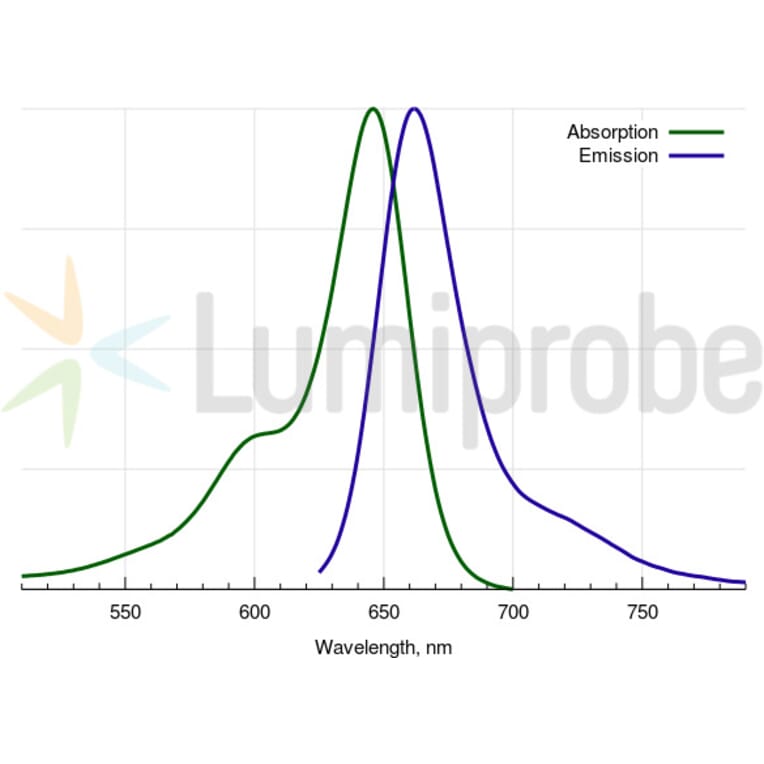
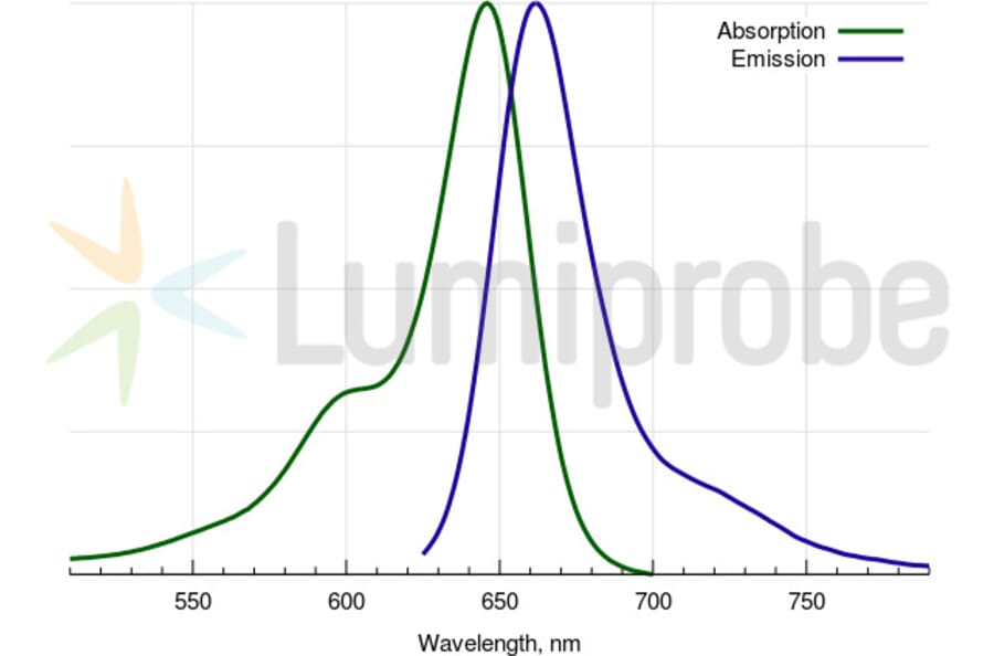
Absorption Maxima
646 nm
Extinction Coefficient
271000 M-1cm-1
Emission Maxima
662 nm
Fluorescence Quantum Yield
0.28
CAS Number
1144107-82-3, 1121756-16-8, 2098639-31-5
CF260
0.04
CF280
0.04
Purity
95% (by 1H NMR and HPLC-MS).
Molecular Formula
C32H37N2KO8S2
Molecular Weight
680,87 Da
Product Form
Dark blue powder.
Solubility
Well soluble in water, DMF, and DMSO (0.35 M = 240 g/L). Practically insoluble in non-polar organic solvents.
Storage
Shipped at room temperature. Upon delivery, store in the dark at -20°C. Avoid prolonged exposure to light.
Disclaimer
This product is for research use only. It is not intended for diagnostic or therapeutic use.