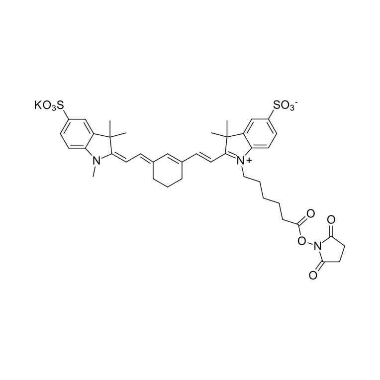
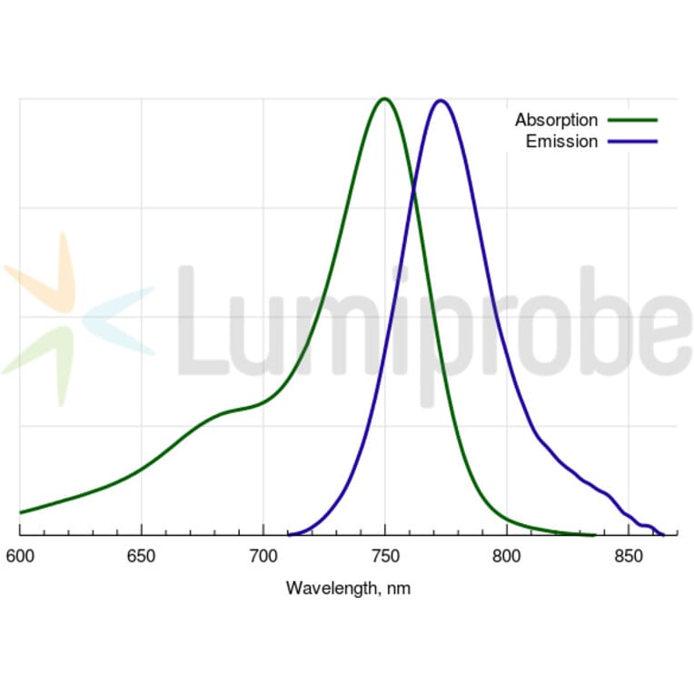
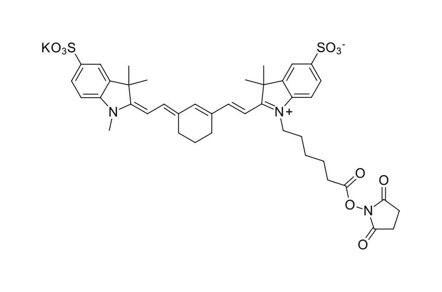
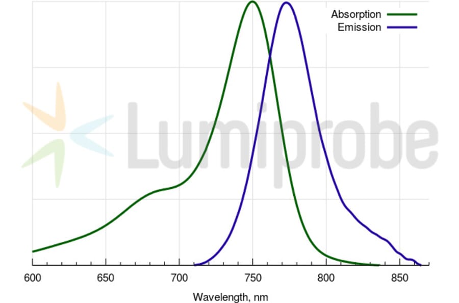
Multimodal imaging, integrating several modalities including down- and up-conversion luminescence, T 1- and T 2(T 2*)-weighted MRI, and CT contrasting in one system, is very promising for improved diagnosis of severe medical disorders. To reach the goal, it is necessary to develop suitable nanoparticles that are highly colloidally stable in biologically relevant media. Here, hydrophilic poly(N,N-dimethylacrylamide-N-acryloylglycine methyl ester)-alendronate-[P(DMA-AGME)-Ale]-coated Gd(Tb)F3:Tb3+(Gd3+),Yb3+,Nd3+ nanoparticles were synthesized by a coprecipitation method in ethylene glycol (EG) followed by coating with the polymer. The particles were tho-roughly characterized by a dynamic light scattering (DLS), transmission electron microscopy (TEM), Fourier-transform infrared spectroscopy (FTIR), thermogravimetric analysis (TGA), X-ray energy dispersive spectroscopy (EDAX), selected area electron diffraction (SAED), elemental ana-lysis and fluorescence spectroscopy. Aqueous particle dispersions exhibited excellent colloidal stability in water and physiological buffers. In vitro toxicity assessments suggested no or only mild toxicity of the surface-engineered Gd(Tb)F3:Tb3+(Gd3+),Yb3+,Nd3+ particles in a wide range of concentrations. Internalization of the particles by several types of cells, including HeLa, HF, HepG2, and INS, was confirmed by a down- and up-conversion confocal microscopy. Newly developed particles thus proved to be an efficient contrast agent for fluorescence imaging, T 1- and T 2(T 2*)-weighted magnetic resonance imaging (MRI), and computed tomography (CT).
Multiple sclerosis (MS) is an autoimmune disease affecting the central nervous system (CNS). Interferon (IFN)-ß constitutes one of the first-line therapies to treat MS, but has limited efficacy due to the injectable systemic administration, short half-life, and limited CNS access. To address these limitations, we developed IFN-ß-loaded chitosan/sulfobutylether-ß-cyclodextrin nanoparticles (IFN-ß-NPs) for delivery of IFN-ß into the CNS via the intranasal (i.n.) route. The nanoparticles (NPs) (˜200 nm, polydispersity ˜0.1, and zeta potential ˜20 mV) were prepared by mixing two aqueous solutions and associated human or murine IFN-ß with high efficiency (90%). Functional in vitro assays showed that IFN-ß-NPs were safe and that IFN-ß was steadily released while retaining biological activity. Biodistribution analysis showed an early and high fluorescence in the brain after nasal administration of fluorescent probe-loaded NPs. Remarkably, mice developing experimental autoimmune encephalomyelitis (EAE), an experimental model of MS, exhibited a significant improvement of clinical symptoms in response to intranasal IFN-ß-NPs (inIFN-ß-NPs), whereas a similar dose of intranasal or systemic free IFN-ß had no effect. Importantly, inIFN-ß-NPs treatment was equally effective despite a reduction of 78% in the total amount of weekly administered IFN-ß. Spinal cords obtained from inIFN-ß-NPs-treated EAE mice showed fewer inflammatory foci and demyelination, lower expression of antigen-presenting and costimulatory proteins on CD11b+ cells, and lower astrocyte and microglia activation than control mice. Therefore, IFN-ß treatment at tested doses was effective in promoting clinical recovery and control of neuroinflammation in EAE only when associated with NPs. Overall, inIFN-ß-NPs represent a potential, effective, non-invasive, and low-cost therapy for MS.