3-color TSA amplification kit for multiplex immunohistochemistry and immunofluorescence using HRP-conjugated antibodies in standard multiplex staining workflows.
Recommended tyramide dye working dilution range: 1:50–1:200 depending on desired signal intensity and optimization requirements.
Assay Type
Tyramide Signal Amplification Kit
Principle of Assay
Tyramide signal amplification (TSA) is a highly sensitive fluorescent method for amplifying immunohistochemical staining when using HRP-conjugated antibodies. After applying HRP-labeled antibody to the sample and washing as in a normal IHC workflow, the tyramide fluorochrome is diluted in tyramide amplification buffer and applied to the samples for 10 minutes to develop the fluorescence. This is then repeated for multiple HRP-labeled antibodies by quenching HRP (HRP-block provided in StreptaClick® HRP Multiplex IHC Kit) and repeating the process for each antibody in sequence.
Shipped at +4°C. Store in the dark at -20°C. Before use, dissolve each vial in 100µl DMSO + 100µl PBS. Aliquot in vials and store at -20°C. The aliquots can be stored in the freezer for at least 12 months. Once thawed, store at +4°C and use within a week.
General Notes
Tyramide dyes should be aliquoted after reconstitution to avoid repeated freeze-thaw cycles.
Disclaimer
This product is for research use only. It is not intended for diagnostic or therapeutic use.
Immunohistochemistry - Tyramide Signal Amplification Kit - 3 Color (A329042)
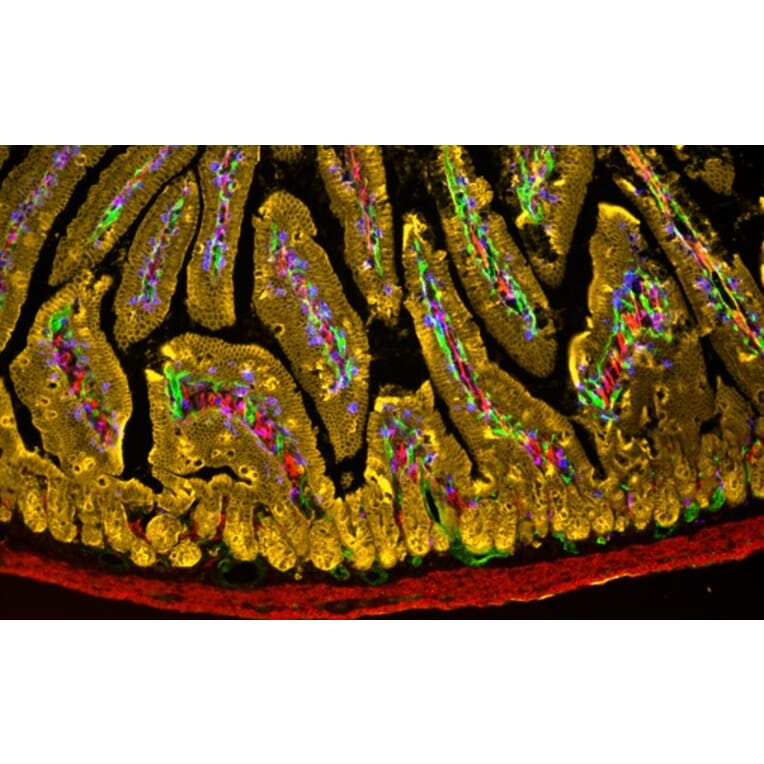
Four-color TSA immunohistochemistry of PFA-fixed, frozen mouse small intestine section using antibodies that have been biotinylated and labelled with the StreptaClick® HRP Complete Tyramide Signal Amplification and Biotinylation Kit - 5 Color. Tissue was mounted in OCT compound and frozen in liquid nitrogen before cutting 10 µm sections. The frozen tissue section was fixed in acetone (30 seconds in ice cold 50% acetone/50%water + 10 min in ice cold 100% acetone), air dried, and washed in PBS. Immunohistochemistry was performed using the following biotinylated antibodies: biotin rat anti-CD31 [MEC13.3] (Tyramide 488; green), biotin mouse anti-human alpha actin [1A4] (Tyramide 594; red), biotin rat anti-CD326 [G8.8] (Tyramide 555; yellow), biotin rat anti-mouse CD8a [53-6.7] (Tyramide 647; blue).
Immunohistochemistry - Tyramide Signal Amplification Kit - 3 Color (A329042)
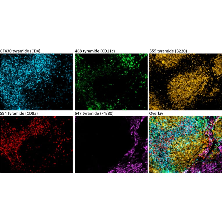
Five-color TSA immunohistochemistry of PFA-fixed, frozen mouse spleen section using antibodies that have been biotinylated and labelled with the StreptaClick® HRP Complete Tyramide Signal Amplification and Biotinylation Kit - 5 Color. Tissue was fixed in 4% paraformaldehyde/10% sucrose for 30 min at +20°C, followed by incubation in 30% sucrose at +4°C overnight before freezing and cutting 10 µm sections. The frozen tissue section was fixed in acetone at room temperature for 10 min, air dried, and washed in PBS. Immunohistochemistry was performed using biotinylated antibodies and TSA dyes for color development: biotin rat anti-mouse CD4 [GK1.5] (Tyramide 430; cyan), biotin hamster anti-mouse CD11c [N418] (Tyramide 488; green), biotin rat anti-mouse B220 [RA3-6B2] (Tyramide 555; yellow), biotin rat anti-mouse CD8a [53-6.7] (Tyramide 594; red), and biotin rat anti-mouse F4/80 [M1/70] (Tyramide 647; magenta).
Publishing research using Tyramide Signal Amplification Kit - 3 Color (A329042)? Please let us know so that we can list the citation on this page.