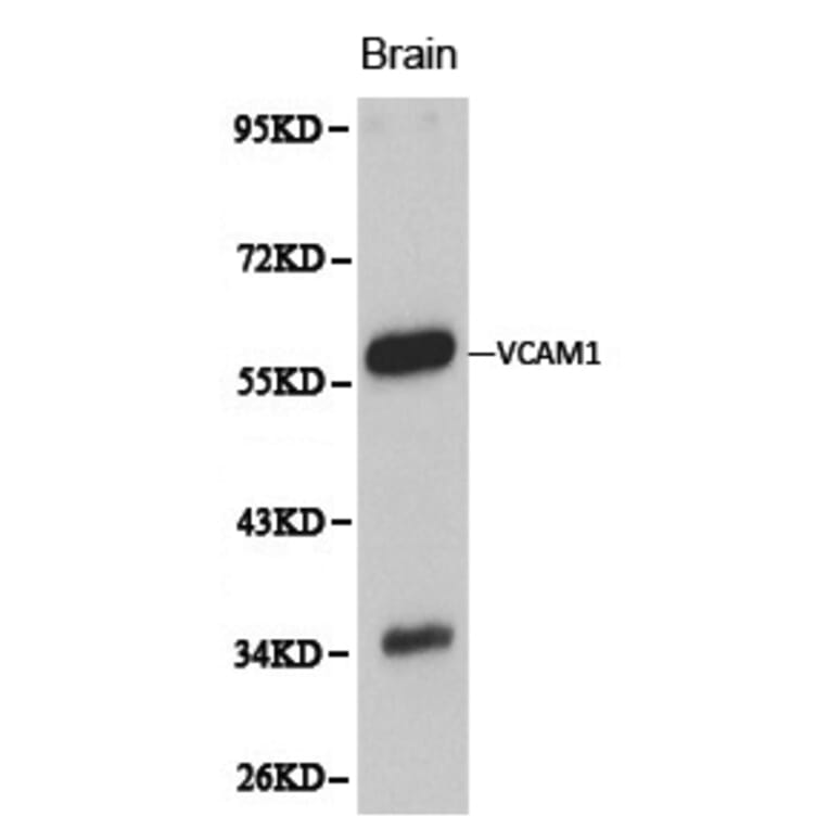
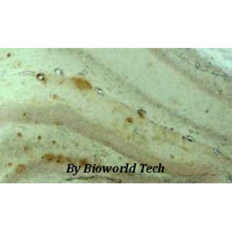
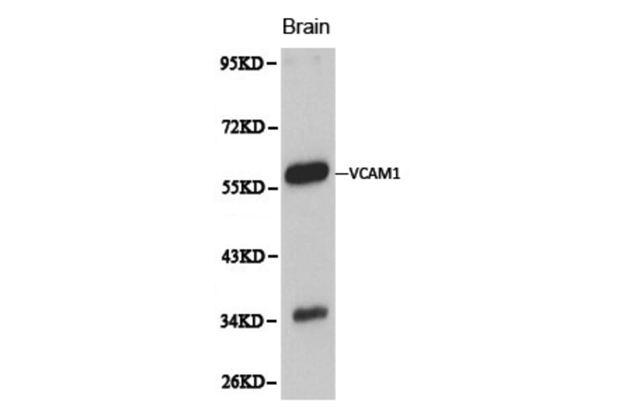
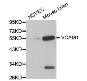
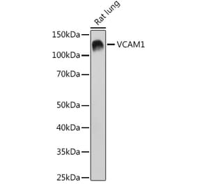
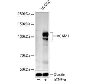
VCAM1 pAb detects endogenous levels of VCAM1 protein.
Applications
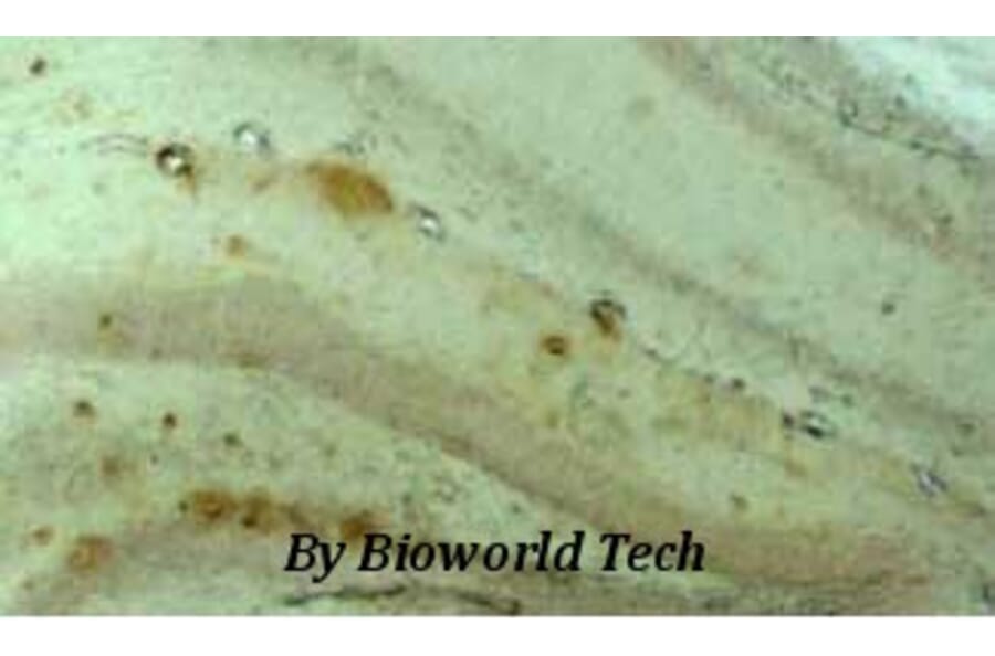
WB, IHC
Reactivity
Human, Mouse, Rat
Immunogen
Recombinant full length Human VCAM1.
Host
Rabbit
Clonality
Polyclonal
Conjugate
Unconjugated
Molecular Weight
~ 58, 81 kDa
Purity
The antibody was affinity-purified from rabbit antiserum by affinity-chromatography using epitope-specific immunogen and the purity is > 95% (by SDS-PAGE).
Product Form
1mg/ml in PBSwith0.1%SodiumAzide,50%Glycerol.
Synonyms
CD106, INCAM-100, V-CAM 1, Vascular cell adhesion protein 1, VCAM-1


![SDS-PAGE - Anti-VCAM1 Antibody [Research Grade Biosimilar] - Low endotoxin, Azide free (A324287) - Antibodies.com](https://cdn.antibodies.com/image/catalog/324/A324287_1.jpg?profile=product_alternative)
![Flow Cytometry - Anti-VCAM1 Antibody [STA] (A86142)](https://cdn.antibodies.com/image/catalog/86/A86142_1.jpg?profile=product_alternative)
![Flow Cytometry - Anti-VCAM1 Antibody [429 (MVCAM.A)] - BSA and Azide free (A121765)](https://cdn.antibodies.com/image/catalog/121/A121765_1.jpg?profile=product_alternative)
![Flow Cytometry - Anti-VCAM1 Antibody [429 (MVCAM.A)] (A121772)](https://cdn.antibodies.com/image/catalog/121/A121772_1.jpg?profile=product_alternative)
![SDS-PAGE - Anti-VCAM1 Antibody [1.4C3] - BSA and Azide free (A250284) - Antibodies.com](https://cdn.antibodies.com/image/catalog/253/A253464_1.jpg?profile=product_alternative)
![SDS-PAGE - Anti-VCAM1 Antibody [1.4C3] (A253461) - Antibodies.com](https://cdn.antibodies.com/image/catalog/250/A250284_1.jpg?profile=product_alternative)
![SDS-PAGE - Anti-VCAM1 Nanobody [cAbVCAM1-5] (A338287) - Antibodies.com](https://cdn.antibodies.com/image/catalog/338/A338287_1.jpg?profile=product_alternative)