Unconjugated
Musk has been traditionally used in East Asia to alleviate the symptoms of angina pectoris. However, it remains unclear as to whether muscone, the main active ingredient of musk, has any beneficial effects on persistent myocardial ischemia in vivo. The aim of the present study was to investigate whether muscone can improve cardiac function and attenuate myocardial remodeling following myocardial infarction (MI) in mice. Mice were subjected to permanent ligation of the left anterior descending coronary artery to induce MI, and then randomly treated with muscone (2 mg/kg/day) or the vehicle (normal saline) for 3 weeks. Sham-operated mice were used as controls and were also administered the vehicle (normal saline). Treatment with muscone significantly improved cardiac function and exercise tolerance, as evidenced by the decrease in the left ventricular end-systolic diameter, left ventricular end-diastolic diameter, as well as an increase in the left ventricular ejection fraction, left ventricular fractional shortening and time to exhaustion during swimming. Pathological and morphological assessments indicated that treatment with muscone alleviated myocardial fibrosis, collagen deposition and improved the heart weight/body weight ratio. Muscone inhibited the inflammatory response by reducing the expression of transforming growth factor (TGF)‑β1, tumor necrosis factor (TNF)-α, interleukin (IL)-1β and nuclear factor (NF)-κB. Treatment with muscone also reduced myocardial apoptosis by enhancing Bcl-2 and suppressing Bax expression. Muscone also induced the phosphorylation of protein kinase B (Akt) and endothelial nitric oxide synthase (eNOS). Our results demonstrate that muscone ameliorates cardiac remodeling and dysfunction induced by MI by exerting anti-fibrotic, anti-inflammatory and anti-apoptotic effects in the ischemic myocardium.
The present study evaluated whether the magnetic nanoparticles of Fe(3)O(4) (MNPs-Fe(3)O(4)) could enhance the activity of artesunate (ART), and to explore its potential mechanisms. Cytotoxicity of the copolymer of ART with MNPs-Fe(3)O(4) on K562 cells was detected by MTT assay and the apoptosis rate of K562 cells was measured by flow cytometry. Protein expression levels of bcl-2, bax, bcl-rambo, caspase-3, and survivin in K562 cells were measured by Western blot. After being incubated with the copolymer of ART with MNPs-Fe(3)O(4) for 48 hours, the growth inhibition rate of K562 cells was significantly increased compared with that of K562 cells treated with ART alone (P < 0.05), and the apoptosis rate of K562 cells was increased significantly compared with that of K562 cells treated with ART alone, suggesting that MNPs-Fe(3)O(4) can enhance the activity of ART. Interestingly, the copolymer-induced cell death was attenuated by caspase inhibitor Z-VAD-FMK. Our results also showed that treatment with the copolymer of MNPs-Fe(3)O(4) and ART increased the expression of bcl-2, bax, bcl-rambo, and caspase-3 proteins, and decreased the expression of survivin protein in K562 cells compared with ART treatment alone. These results suggest that MNPs-Fe(3)O(4) can enhance ART-induced apoptosis, which may be related to the upregulation of bcl-rambo and downregulation of survivin.


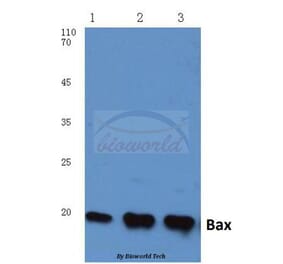
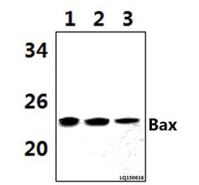
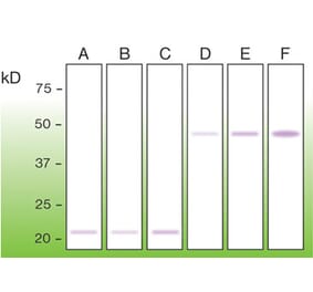
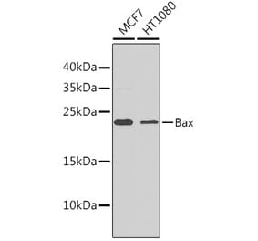
![Western Blot - Anti-Bax Antibody [ARC5006-10] (A306010) - Antibodies.com](https://cdn.antibodies.com/image/catalog/306/A306010_1.jpg?profile=product_alternative)
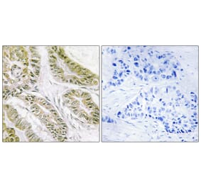
![Immunohistochemistry - Anti-Bax Antibody [2D2] (A249822) - Antibodies.com](https://cdn.antibodies.com/image/catalog/249/A249822_1.jpg?profile=product_alternative)

![Immunohistochemistry - Anti-Bax Antibody [2D2] - BSA and Azide free (A253002) - Antibodies.com](https://cdn.antibodies.com/image/catalog/253/A253002_1.jpg?profile=product_alternative)
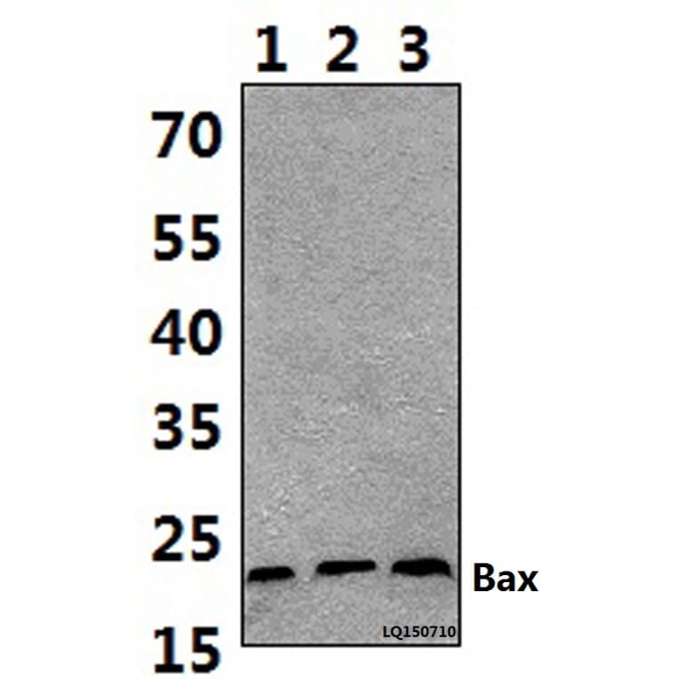
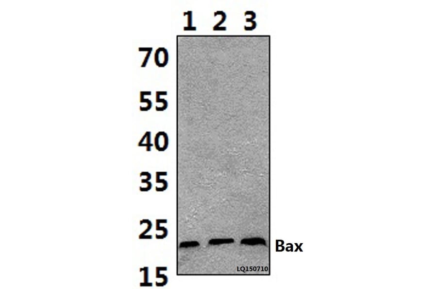
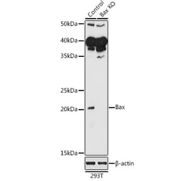
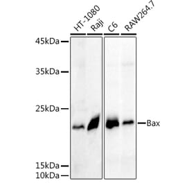
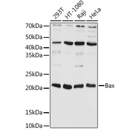
![Western Blot - Anti-Bax Antibody [ARC0164] (A307133) - Antibodies.com](https://cdn.antibodies.com/image/catalog/307/A307133_1.jpg?profile=product_alternative)