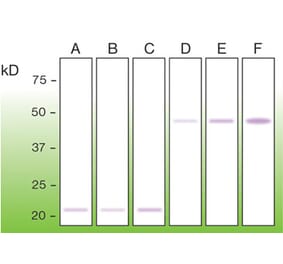
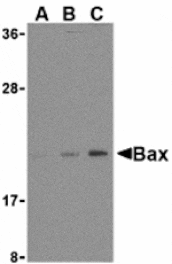
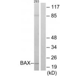
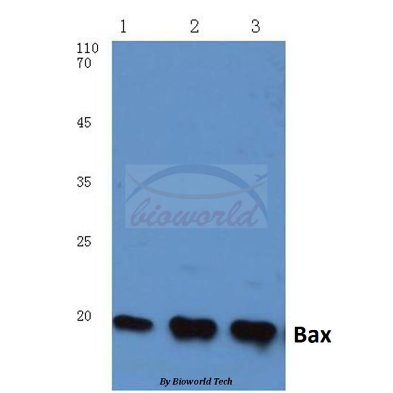
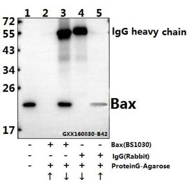
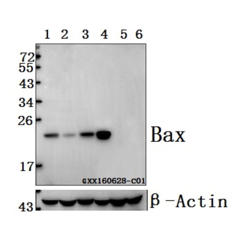
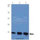
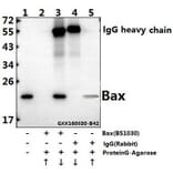
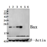
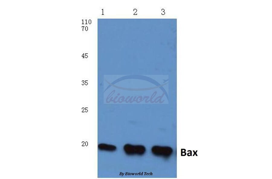
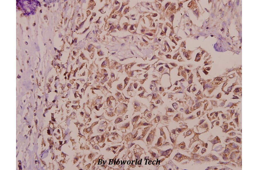
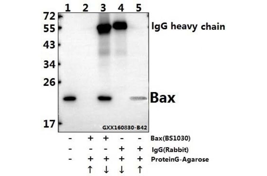
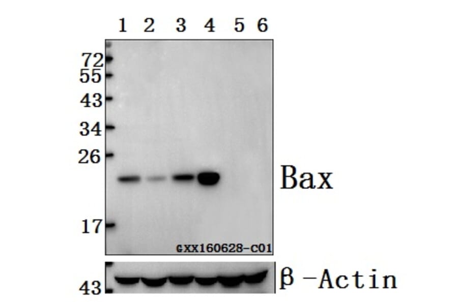
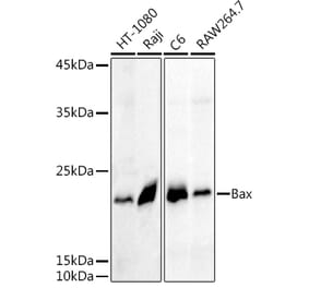
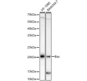
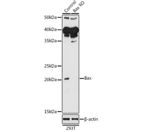
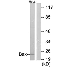
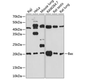
Bax (S4) pAb detects endogenous levels of Bax protein.
Applications
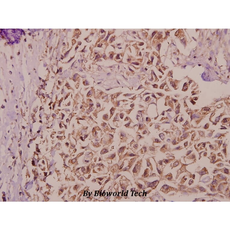
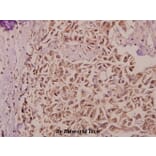
WB, IHC
Reactivity
Human, Mouse, Rat
Immunogen
Synthetic peptide, corresponding to the N-terminus of Human Bax.
Host
Rabbit
Clonality
Polyclonal
Conjugate
Unconjugated
Molecular Weight
~ 21 kDa
Purity
The antibody was affinity-purified from rabbit antiserum by affinity-chromatography using epitope-specific immunogen and the purity is > 95% (by SDS-PAGE).
Product Form
1 mg/ml in Phosphate buffered saline (PBS) with 0.05% sodium azide, approx. pH 7.2.
Synonyms
Apoptosis regulator BAX, Bcl-2-like protein 4, Bcl2-L-4, BCL2L4


![Immunohistochemistry - Anti-Bax Antibody [2D2] (A278397) - Antibodies.com](https://cdn.antibodies.com/image/catalog/249/A249822_1.jpg?profile=product_alternative)

![Immunohistochemistry - Anti-Bax Antibody [2D2] - BSA and Azide free (A249822) - Antibodies.com](https://cdn.antibodies.com/image/catalog/253/A253002_1.jpg?profile=product_alternative)