Mouse monoclonal [4A7] antibody to Aurora A + B + C Kinase.
Specificity
This antibody was raised against human Aurora A protein and was shown to also bind Aurora B and Aurora C; as a result the epitope is likely within the highly conserved serine/threonine kinase domain. This antibody can be used to identify dividing or soon to be dividing cells and is also an excellent marker of midbodies both during and after cell division.
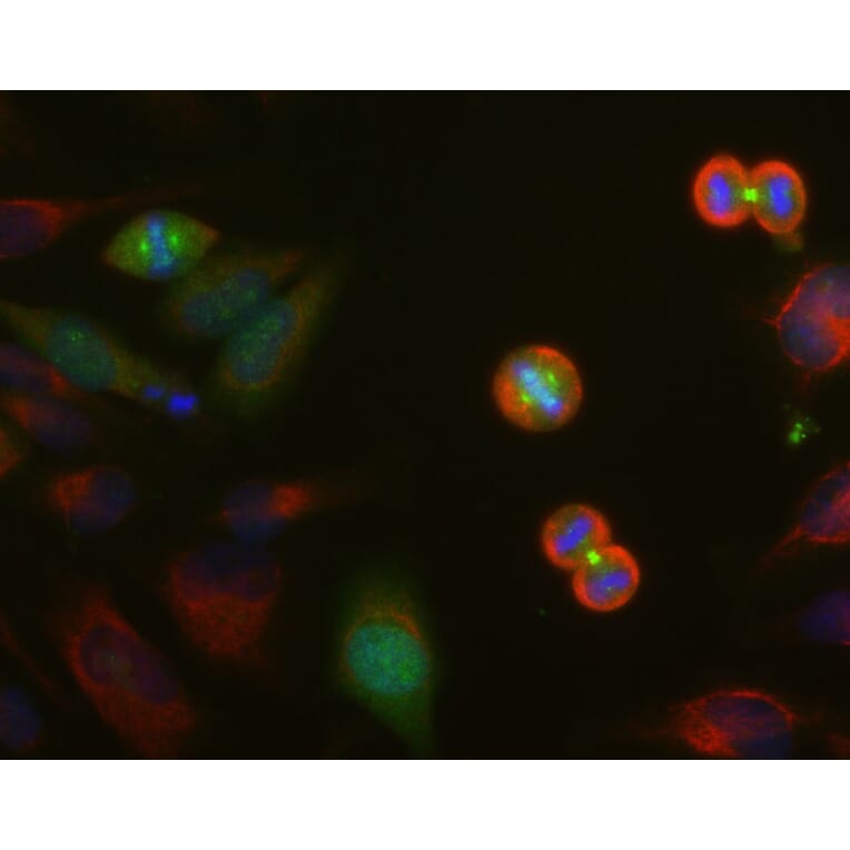
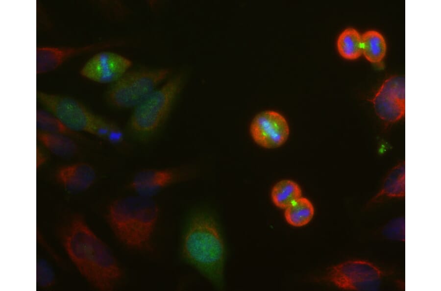
Immunofluorescence - Anti-Aurora A + B + C Kinase Antibody [4A7] (A85401)
HeLa cell cultures were stained with Anti-Aurora A/B/C Kinase Antibody (green). In this image, strong staining in spindle poles was seen in cells at anaphase. This antibody also stains the midbodies between the two daughter cells. Cells were counterstained with Anti-Vimentin Antibody (A85421 | red). Blue is a DNA stain.
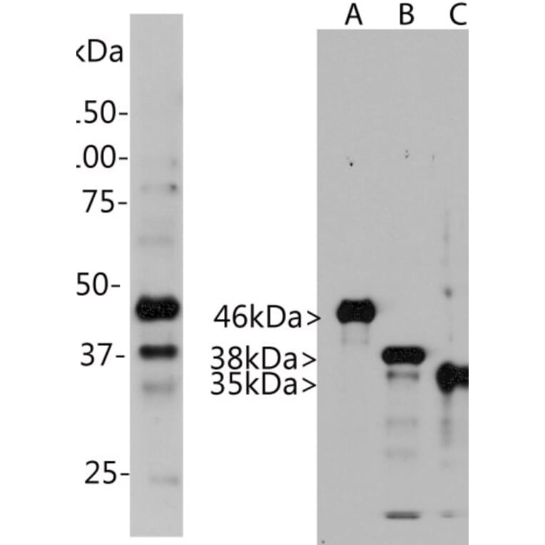
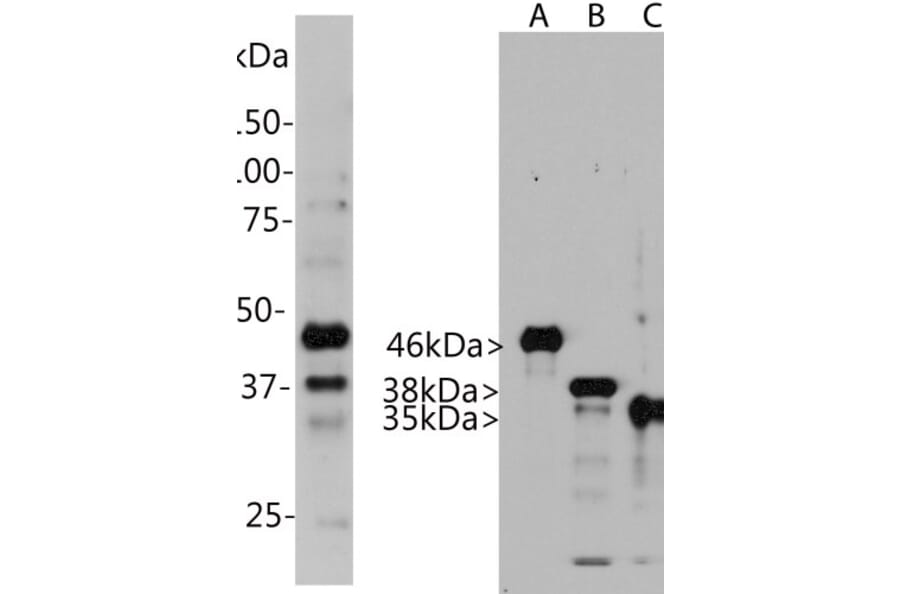
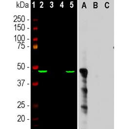
Western Blot - Anti-Aurora A + B + C Kinase Antibody [4A7] (A85401)
Left: Western blot analysis of Anti-Aurora A/B/C Kinase Antibody in HeLa cells. Blot of HeLa cells treated with 100ng/ml nocodazole for 18 hours was probed with Anti-Aurora A/B/C Kinase Antibody. Nocodazole is a microtubule depolymerizating agent which induces cells to halt at the G2/M phase and also induces Aurora kinase expression. The Anti-Aurora A/B/C Kinase Antibody binds strongly to bands at about 46 kDa and 38kDa, corresponding to Aurora A and Aurora B. Also, it recognizes a weak band at 35 kDa which is Aurora C. Right: Blot of recombinant full length human Aurora A, B and C was probed with Anti-Aurora A/B/C Kinase Antibody. This antibody reacts strongly with all three Aurora kinases proteins.
Alternative products to Anti-Aurora A + B + C Kinase Antibody [4A7] (A85401)