Section of rat hippocampus stained with Anti-c-Fos Antibody (red) and Anti-FOX3/NeuN Antibody (A85403 | green). DAPI reveals nuclei of neurons and glia in blue. The hippocampal neurons stain green for FOX3/NeuN and a few also are expressing c-FOS, and so appear orange. These cells were spontaneously active at the time the animal was sacrificed.
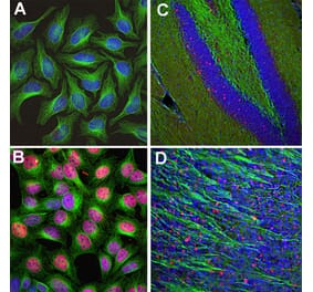
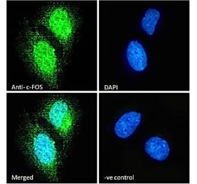
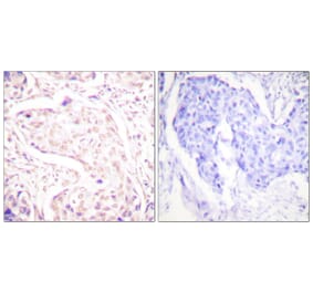
Left: Anti-c-Fos Antibody staining (green) in HeLa cells, which were treated with serum-starvation for 36 hours, followed by 2 hours, 20% FBS stimulation (bottom panel), or PBS treatment (top panel). Green c-Fos staining only localizes in the nuclei of stimulated cells, but not in un-stimulated cells. Cells are counter-stained with Anti-Vimentin Antibody (A85421 | red). Blue shows DAPI staining of nucleus. Middle: Mouse brain section (45 µM; fixed by transcardial perfusion with 4% paraformaldehyde) labeled with Anti-c-Fos Antibody using a standard HRP-DAB (horseradish peroxidase-3,3’-diaminobenzidine) staining technique. Cells expressing c-Fos show dark color in nucleus. Right: Mouse cortical section labeled with Anti-c-Fos Antibody (red) and Anti-Fox3/NeuN Antibody (A85403 | green) using immuno-fluorescent confocal-microscopy. Neurons positive for c-Fos and Fox3/NeuN appear to be yellow. Inset shows an enlarged image of Anti-c-Fos Antibody staining. Nuclei are labeled with DAPI (blue).
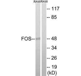
Western blot analysis of cell lysates using Anti-c-Fos Antibody [2H2] (A85387), at a dilution of 1:1,000, in green, and Anti-GAPDH Antibody (A85377), at a dilution of 1:20,000, in red, used as a loading control. The lanes contain samples of: [Lane 1] Protein standards, in red, [Lane 2] HeLa cells in serum free media, [Lane 3] HeLa cells stimulated with 20% fetal bovine serum for 2hrs after 36hrs in serum free media, [Lane 4] rat cortical neurons, and [Lane 5] rat cortical neurons treated with membrane depolarization buffer for 5hrs. Multiple bands at 50-65kDa in stimulated or treated cell lysates correspond to different forms of the c-Fos proten. The single band at 37 kDa represents GAPDH protein.
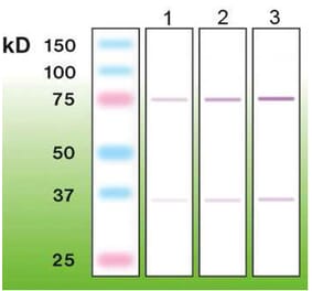
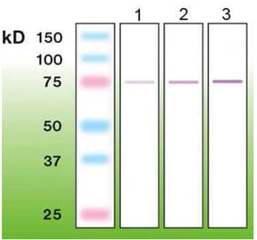
Binding curve set for Anti-c-Fos Antibody [2H2] (A85387) with 25nM IgG and limiting dilutions of recombinant c-FOS protein (0-350nM) obtained using label-free bio-layer interferometry system (Octet RED96). Color-coded traces show sensorgram data normalized to baseline after subtraction of 0nM IgG signal from all channels. Traces with overlying fit lines in red indicate their inclusion in the global fit analysis used to derive kinetic parameters listed under the legend (R^2 – goodness of correlation between the fit and data; kon – association rate constant; koff – dissociation rate constant; KD = koff/kon – affinity constant/equilibrium dissociation constant).
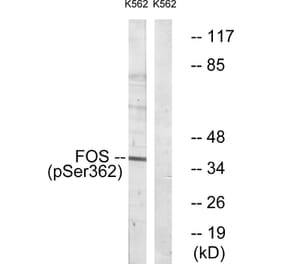
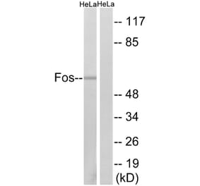
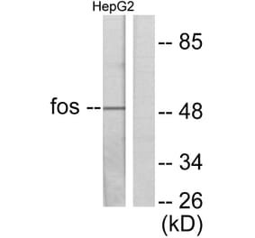
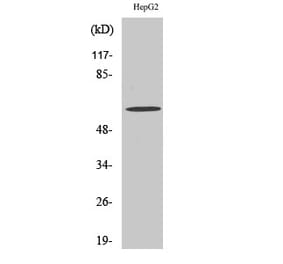
Top panel: Western blot analysis of c-Fos expression in HeLa cells using Anti-c-Fos Antibody. Lane 1: HeLa cells were serum-starved for 36 hours. 2: Serum-starved HeLa cells were stimulated with 20% FBS (fetal bovine serum) for 2 hours. Anti-c-Fos Antibody recognizes bands in the range of 50-65 kDa, which represent multiple forms of c-Fos. Serum starvation attenuates c-Fos expression, while 20% FBS strongly stimulates c-Fos expression. Bottom panel: Blot was stripped and probed with Anti-GAPDH Antibody (A85382), used as loading control.
Rat brain neural cultures stimulated with membrane deplorization buffer for 5 hours. This is a salt solution containing 170mM Potassium which depolarizes and stimulates gene expression in neuronal cells but has no effect on glia. Cultures were stained with Anti-c-Fos Antibody [2H2] (A85387) (green) and Anti-GFAP Antibody (A85419) (red). Nuclei were stained with DAPI (blue).
Rat brain neural cultures. This is a salt solution containing 170mM Potassium which depolarizes and stimulates gene expression in neuronal cells but has no effect on glia. Cultures were stained with Anti-c-Fos Antibody [2H2] (A85387) (green) and Anti-GFAP Antibody (A85419) (red). Nuclei were stained with DAPI (blue).
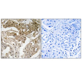
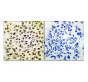
Immunohistochemistry analysis of formalin Fixed Paraffin Embedded Mouse Material. Mouse hippocampus was stained with Anti-c-Fos Antibody [2H2] (A85387) at a dilution of 1:1,000 detected in DAB (brown) following the Vector Labs Mouse on Mouse ImmPRESS® HRP method. Counterstained with Hematoxylin (blue). Anti-c-Fos Antibody [2H2] (A85387) specifically detects the nuclei of a few spontaneously active neurons.
Immunohistochemistry analysis of formalin Fixed Paraffin Embedded Rat Material. Rat hippocampus was stained with Anti-c-Fos Antibody [2H2] (A85387) at a dilution of 1:1,000 detected with DAB (brown) using the Vector Labs ImmPRESS method and reagents with citra buffer retrieval. Counterstained with Hematoxylin (blue). Anti-c-Fos Antibody [2H2] (A85387) specifically detects the nuclei of spontaneously active or experimentally stimulated neurons.


![Immunofluorescence - Anti-c-Fos Antibody [2H2] (A85387) - Antibodies.com](https://cdn.antibodies.com/image/catalog/85/A85387_1.jpg?profile=product_top)
![Immunofluorescence - Anti-c-Fos Antibody [2H2] (A85387) - Antibodies.com](https://cdn.antibodies.com/image/catalog/85/A85387_2.jpg?profile=product_top)
![Western Blot - Anti-c-Fos Antibody [2H2] (A85387) - Antibodies.com](https://cdn.antibodies.com/image/catalog/85/A85387_3.jpg?profile=product_top)
![Binding curve - Anti-c-Fos Antibody [2H2] (A85387) - Antibodies.com](https://cdn.antibodies.com/image/catalog/85/A85387_4.jpg?profile=product_top)
![Western Blot - Anti-c-Fos Antibody [2H2] (A85387) - Antibodies.com](https://cdn.antibodies.com/image/catalog/85/A85387_5.jpg?profile=product_top)
![Immunofluorescence - Anti-c-Fos Antibody [2H2] (A85387) - Antibodies.com](https://cdn.antibodies.com/image/catalog/85/A85387_6.jpg?profile=product_top)
![Immunofluorescence - Anti-c-Fos Antibody [2H2] (A85387) - Antibodies.com](https://cdn.antibodies.com/image/catalog/85/A85387_7.jpg?profile=product_top)
![Immunohistochemistry - Anti-c-Fos Antibody [2H2] (A85387) - Antibodies.com](https://cdn.antibodies.com/image/catalog/85/A85387_8.jpg?profile=product_top)
![Immunohistochemistry - Anti-c-Fos Antibody [2H2] (A85387) - Antibodies.com](https://cdn.antibodies.com/image/catalog/85/A85387_9.jpg?profile=product_top)
![Immunofluorescence - Anti-c-Fos Antibody [2H2] (A85387) - Antibodies.com](https://cdn.antibodies.com/image/catalog/85/A85387_1.jpg?profile=product_top_thumb)
![Immunofluorescence - Anti-c-Fos Antibody [2H2] (A85387) - Antibodies.com](https://cdn.antibodies.com/image/catalog/85/A85387_2.jpg?profile=product_top_thumb)
![Western Blot - Anti-c-Fos Antibody [2H2] (A85387) - Antibodies.com](https://cdn.antibodies.com/image/catalog/85/A85387_3.jpg?profile=product_top_thumb)
![Binding curve - Anti-c-Fos Antibody [2H2] (A85387) - Antibodies.com](https://cdn.antibodies.com/image/catalog/85/A85387_4.jpg?profile=product_top_thumb)
![Western Blot - Anti-c-Fos Antibody [2H2] (A85387) - Antibodies.com](https://cdn.antibodies.com/image/catalog/85/A85387_5.jpg?profile=product_top_thumb)
![Immunofluorescence - Anti-c-Fos Antibody [2H2] (A85387) - Antibodies.com](https://cdn.antibodies.com/image/catalog/85/A85387_6.jpg?profile=product_top_thumb)
![Immunofluorescence - Anti-c-Fos Antibody [2H2] (A85387) - Antibodies.com](https://cdn.antibodies.com/image/catalog/85/A85387_7.jpg?profile=product_top_thumb)
![Immunofluorescence - Anti-c-Fos Antibody [2H2] (A85387) - Antibodies.com](https://cdn.antibodies.com/image/catalog/85/A85387_1.jpg?profile=product_image)
![Immunofluorescence - Anti-c-Fos Antibody [2H2] (A85387) - Antibodies.com](https://cdn.antibodies.com/image/catalog/85/A85387_2.jpg?profile=product_image)
![Western Blot - Anti-c-Fos Antibody [2H2] (A85387) - Antibodies.com](https://cdn.antibodies.com/image/catalog/85/A85387_3.jpg?profile=product_image)
![Binding curve - Anti-c-Fos Antibody [2H2] (A85387) - Antibodies.com](https://cdn.antibodies.com/image/catalog/85/A85387_4.jpg?profile=product_image)
![Western Blot - Anti-c-Fos Antibody [2H2] (A85387) - Antibodies.com](https://cdn.antibodies.com/image/catalog/85/A85387_5.jpg?profile=product_image)
![Immunofluorescence - Anti-c-Fos Antibody [2H2] (A85387) - Antibodies.com](https://cdn.antibodies.com/image/catalog/85/A85387_6.jpg?profile=product_image)
![Immunofluorescence - Anti-c-Fos Antibody [2H2] (A85387) - Antibodies.com](https://cdn.antibodies.com/image/catalog/85/A85387_7.jpg?profile=product_image)
![Immunohistochemistry - Anti-c-Fos Antibody [2H2] (A85387) - Antibodies.com](https://cdn.antibodies.com/image/catalog/85/A85387_8.jpg?profile=product_image)
![Immunohistochemistry - Anti-c-Fos Antibody [2H2] (A85387) - Antibodies.com](https://cdn.antibodies.com/image/catalog/85/A85387_9.jpg?profile=product_image)