ISO 9001:2015 Certified
Live Customer Support
4.5/5 on Trustpilot
100% Quality Guarantee

Anti-CD4 Antibody [CA13.1E4] (A279972)

$560

Mouse monoclonal [CA13.1E4] antibody to CD4 for IHC-Fr, IP and Flow Cytometry.

Shipping Information

$40
Dispatched from St. Louis, MO.
Lead Time: 5-7 business days.
Name
Anti-CD4 Antibody [CA13.1E4]
Description
Mouse monoclonal [CA13.1E4] antibody to CD4.
Specificity
This antibody recognises a population of CD8 positive cells in the canine thymus. CD4 expression has also been reported on a population of CD3 positive peripheral blood T lymphocytes which are double positive for CD4 and CD8 (Bismarck et al. 2012 ). Similar reports have been made for cynomolgus monkey , pig (Saalmuller et al. 1987 , Pescowitz et al. 1990) as well as rat, chicken and human (reviewed Zuckermann 1999 ).Uniquely amongst mammalian species clone CA13.1E4 also recognises CD4 expressed on canine neutrophils at a similar density to that expressed on canine T helper cells. The functional significance of this is not understood and remains enigmatic in view of the roles described for CD4 in canine and other mammalian species. Clone CA13.1E4 also demonstrates immunohistological staining of splenic marginal zone macrophages. Other macrophage populations, such as splenic red pulp macrophages and Langerhans cells do not stain with clone CA13.1E4.Canine CD4 positive T cells can be further characterized according to their simultaneous expression of CD45RA as recognized by Rat anti Canine anti CD45RA clone CA4.1D3 in a manner analogous to that seen with human T cells. Clone CA13.1E4 has been used amongst a large panel of anti-canine monoclonal antibodies for the study of various lymphoproliferative diseases. T lymphocytes in large granular lymphocyte (LGL) lymphocytosis are negative for CD4 (McDonough and Moore 2000 ). Dogs with chronic myelogenous leukemia demonstrate CD4 positive staining on neutrophils. CD4 expression varies considerably between various canine leukemias.
Applications
IHC-Fr, IP, Flow Cytometry
Dilutions
Flow Cytometry: Neat, Use 25µl of the suggested working dilution to label 106 cells or 100µl whole blood
Reactivity
Canine
Immunogen
Canine Thymocytes.
Host
Mouse
Clonality
Monoclonal
Clone ID
CA13.1E4
Isotype
IgG1
Conjugate

Unconjugated

Concentration
Lot Specific
Purity
Tissue Culture Supernatant
Product Form
Liquid
Formulation
Supplied in <0.1% Sodium Azide.
Storage
Shipped at ambient temperature. Upon delivery aliquot and store at -20°C. When thawed, aliquot the sample as needed. Short term (up to 4 weeks): store at 4°C. Long term: store at -20°C. Avoid freeze / thaw cycles. Storage in frost free freezers is not recommended.
General Notes
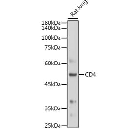
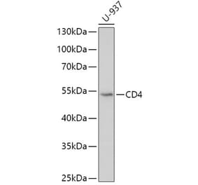
Mouse anti Dog CD4 antibody (CA13.1E4), a monoclonal antibody specific for canine CD4. CA13.1E4 was clustered at the first Canine Leukocyte Antigen Workshop (Claw) [Cobbold et al. 1992 ] and identifies, by immunoprecipitation a ~60 kDa monomeric protein under both reducing and non-reducing conditions. Canine CD4 is a surface glycoprotein expressed by non CD8 expressing T lymphocytes, with similar developmental and functional characteristics to T helper cells described in other mammalian species. Mouse anti Dog CD4 also recognizes a population of CD8 positive cells in the canine thymus (Moore et al. 1992). CD4 expression has also been reported on a population of CD3 positive peripheral blood T lymphocytes which are double positive for CD4 and CD8 (Bismarck et al. 2012 ). Similar reports have been made for cynomolgus monkey (Nam et al. 2000), pig (Saalmuller et al. 1987 , Pescowitz et al. 1990) as well as rat, chicken and human (reviewed Zuckermann 1999 ). Uniquely amongst mammalian species clone CA13.1E4 also recognizes CD4 expressed on canine neutrophils at a similar density to that expressed on canine T helper cells. The functional significance of this is not understood and remains enigmatic in view of the roles described for CD4 in canine and other mammalian species (Moore et al. 1992). Clone CA13.1E4 also demonstrates immunohistological staining of splenic marginal zone macrophages (Moore et al. 1992). Other macrophage populations, such as splenic red pulp macrophages and Langerhans cells do not stain with clone CA13.1E4. Canine CD4 positive T cells can be further characterized according to their simultaneous expression of CD45RA as recognized by Rat anti Canine anti CD45RA clone CA4.1D3 in a manner analogous to that seen with human T cells (Moore et al. 1992). Clone CA13.1E4 has been used amongst a large panel of anti-canine monoclonal antibodies for the study of various lymphoproliferative diseases. T lymphocytes in large granular lymphocyte (LGL) lymphocytosis are negative for CD4 (McDonough and Moore 2000 ). Dogs with chronic myelogenous leukemia demonstrate CD4 positive staining on neutrophils (Tarrant et al. 2001). CD4 expression varies considerably between various canine leukemias.
Synonyms
T-cell surface antigen T4/Leu-3, T-cell surface glycoprotein CD4
Disclaimer
This product is for research use only. It is not intended for diagnostic or therapeutic use.
Publishing research using Anti-CD4 Antibody [CA13.1E4] (A279972)? Please let us know so that we can list the citation on this page.

Alternative products to Anti-CD4 Antibody [CA13.1E4] (A279972)

Proteins predicted to interact with CD4

Predicted protein interactions based upon String database. Revelancy score correlates with probability of interaction.
99.9% Relevancy Score
99.8% Relevancy Score
99.7% Relevancy Score
99.6% Relevancy Score