This antibody targets the C-terminal domain of human CXCR4, recognizing the non-phosphorylated isoform. It is optimized for Western blot analysis, enables immunoprecipitation from brain tissue lysates, supports immunohistochemistry in cultured cells and tissue sections, and exhibits cross-reactivity with human, mouse, and rat species.
Applications
WB, ICC, IHC
Dilutions
WB: 1:1,000, ICC: 1:200, IHC: 1:100
Reactivity
Human, Mouse, Rat
Immunogen
Synthetic peptide corresponding to human, mouse and rat CXCR4 (amino acids. 338-359). Immunogen range is 22-22 amino acids.
Sequence
KGKRGGHSSVSTESESSSFHSS
Host
Rabbit
Clonality
Polyclonal
Isotype
IgG
Conjugate
Unconjugated
Purification
Antigen affinity purification.
Concentration
Lot Specific
Product Form
Liquid
Formulation
Supplied in Dulbecco's PBS, pH 7.4, with 150 mM NaCl and 0.005% Sodium Azide.
Storage
Shipped at 4°C. Upon delivery aliquot and store at -20°C. Avoid freeze/thaw cycles.
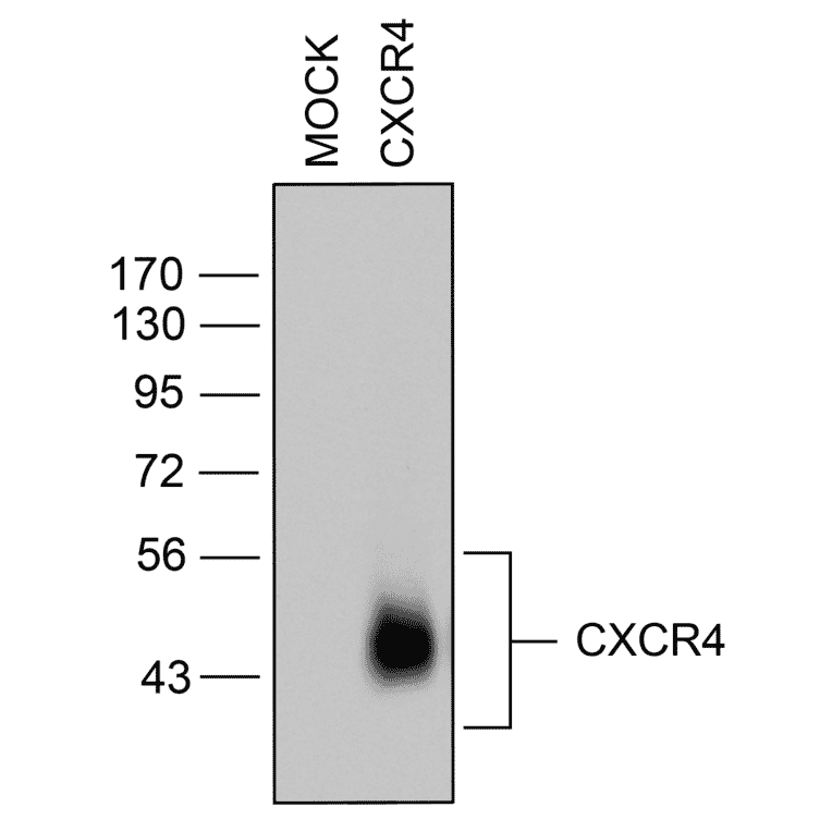
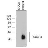
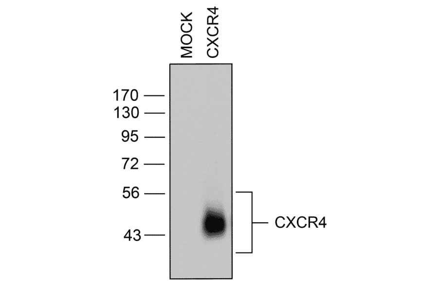
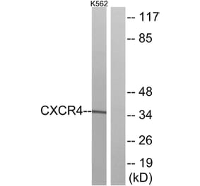
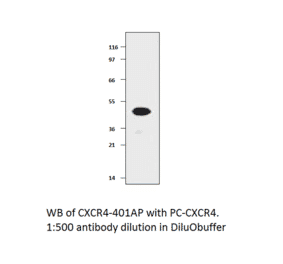
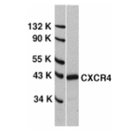
Western blot validation of CXC Chemokine Receptor 4 (CXCR4) in transfected HEK293 cells. Lysates from native HEK293 cells (MOCK) or cells stably expressing CXCR4 were immunoblotted with anti-CXCR4 antibody (A334501) at a 1:1000 dilution.
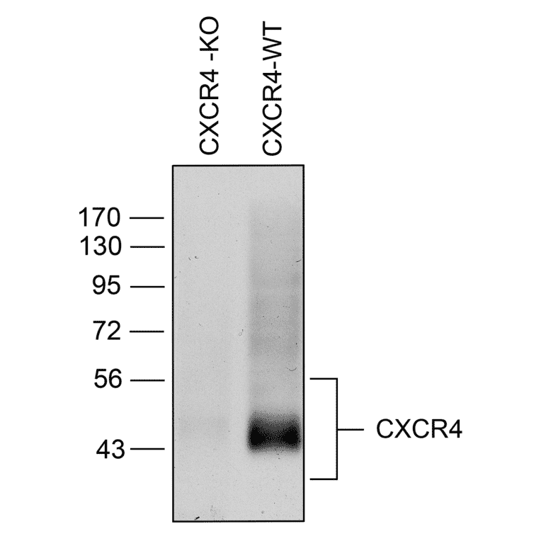
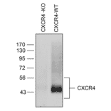
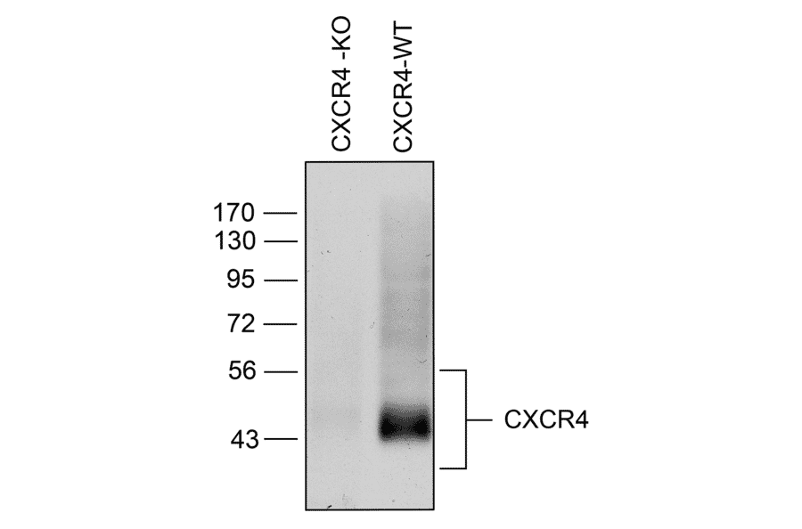
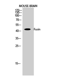
Analysis of CXC Chemokine Receptor 4 (CXCR4) expression in mouse brain by Western blot. Brain homogenates from CXCR4-deficient (CXCR4-KO) and wild-type (CXCR4-WT) mice were immunoblotted with anti-CXCR4 antibody (A334501) at 1:1000. CXCR4 receptors were detected in CXCR4-WT but absent in CXCR4-KO mice.
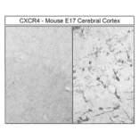
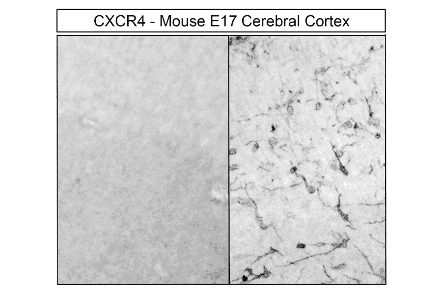
Immunohistochemical localization of CXC Chemokine Receptor 4 (CXCR4) in mouse cerebral cortex. E17 mouse forebrain sections were dewaxed, microwaved in citric acid, and stained with anti-CXCR4 antibody (A334501) at 1:100, followed by biotinylated anti-rabbit IgG and avidin-biotin solution. Sections were developed with 3,3-diaminobenzidine (DAB)-glucose oxidase and counterstained with hematoxylin. CXCR4 receptors were observed at the plasma membrane of tangentially migrating neurons.
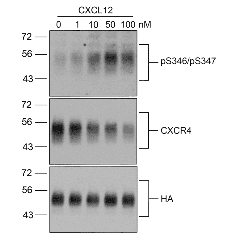
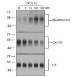
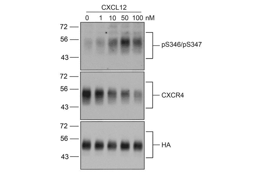
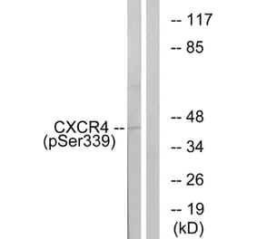
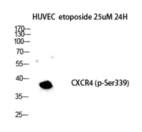
Dose-response analysis of CXC Chemokine Receptor 4 (CXCR4) phosphorylation at Serine346/Serine347. HEK293 cells stably expressing HA-tagged CXCR4 were untreated or treated with 1 nM to 100 nM CXCL12 for 30 min. Lysates were probed with anti-CXCR4 (Phospho Ser346 + Ser347) antibody (A334500) at 1:1000 (upper panel), stripped and reprobed with anti-CXCR4 antibody (A334501) at 1:1000 (middle panel), and stripped again and reprobed with anti-HA antibody at 1:1000 for equal loading (lower panel).
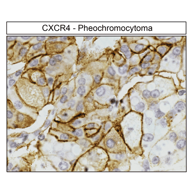
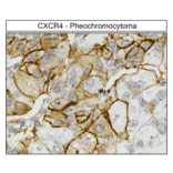
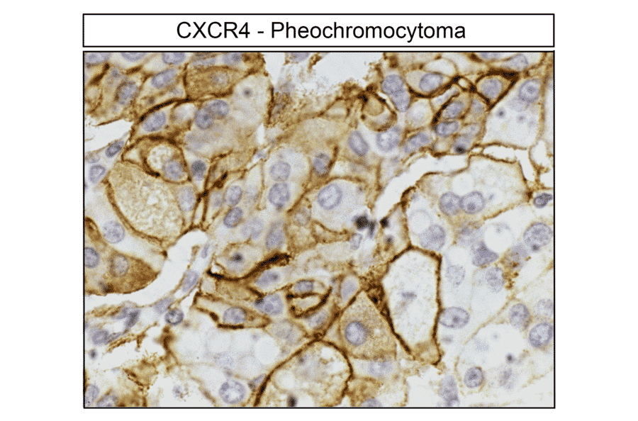
Immunohistochemical detection of CXC Chemokine Receptor 4 (CXCR4) in human pheochromocytoma. Tumor sections were dewaxed, microwaved in citric acid, and incubated with anti-CXCR4 antibody (A334501) at 1:100, followed by biotinylated anti-rabbit IgG and avidin-biotin solution. Sections were developed with 3,3-diaminobenzidine (DAB)-glucose oxidase and counterstained with hematoxylin. CXCR4 receptors were uniformly detected at the plasma membrane of tumor cells.
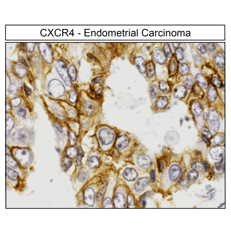
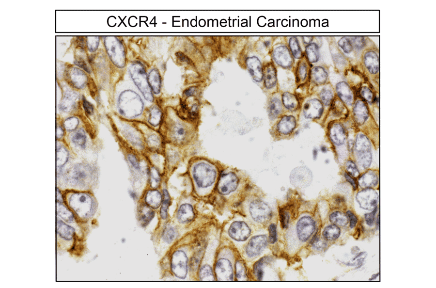
Localization of CXC Chemokine Receptor 4 (CXCR4) in human endometrial carcinoma by immunohistochemistry. Tumor sections were dewaxed, microwaved in citric acid, and stained with anti-CXCR4 antibody (A334501) at 1:100, followed by biotinylated anti-rabbit IgG and avidin-biotin solution. Sections were developed with 3,3-diaminobenzidine (DAB)-glucose oxidase and counterstained with hematoxylin. CXCR4 receptors were uniformly observed at the plasma membrane of tumor cells.
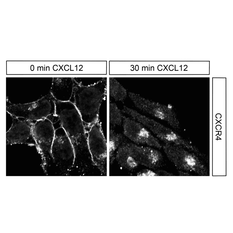
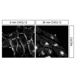
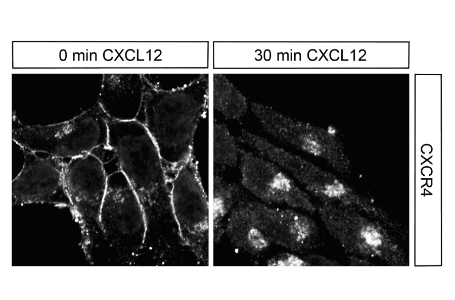
Immunocytochemical analysis of CXC Chemokine Receptor 4 (CXCR4) in HEK293 cells. HEK293 cells stably expressing CXCR4 were untreated or treated with 100 nM CXCL12 for 30 min and stained with anti-CXCR4 antibody (A334501) at 1:200. CXCR4 receptors were localized to the plasma membrane in untreated cells (0 min) and in perinuclear vesicular clusters after 30 min of CXCL12 exposure.
Recombinant human monoclonal antibody to CXCR4 for use as a research grade Ulocuplumab biosimilar for ELISA, Flow Cytometry, Functional Studies and in vivo Research.
Recombinant human monoclonal antibody to CXCR4 for use as a research grade MEDI3185 biosimilar for ELISA, Flow Cytometry, Functional Studies and in vivo Research.
Recombinant human monoclonal antibody to CXCR4 for use as a research grade Anti-Human CXCR4 [CF172] for ELISA, Flow Cytometry, Functional Studies and in vivo Research.


![Flow Cytometry - Anti-CXCR4 Antibody [12G5] (A86617)](https://cdn.antibodies.com/image/catalog/86/A86617_1.jpg?profile=product_alternative)
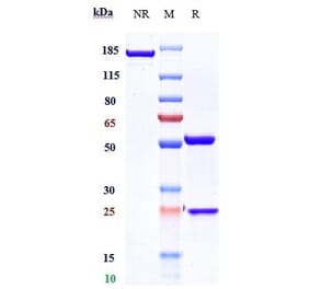
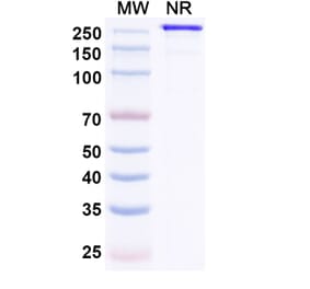
![SDS-PAGE - Anti-CXCR4 Antibody [Research Grade Biosimilar] - Low endotoxin, Azide free (A323988) - Antibodies.com](https://cdn.antibodies.com/image/catalog/323/A323988_1.jpg?profile=product_alternative)
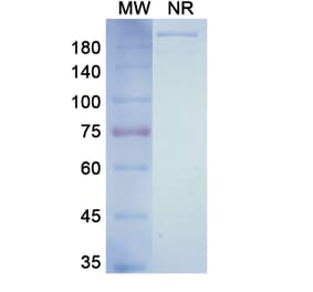
![SDS-PAGE - Anti-CXCR4 Antibody [CF172] Biosimilar - BSA and Azide free (A339648) - Antibodies.com](https://cdn.antibodies.com/image/catalog/339/A339648_1.jpg?profile=product_alternative)