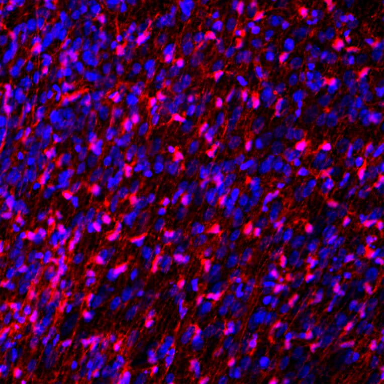
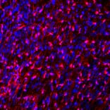
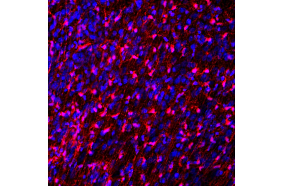
Immunofluorescent analysis of rat embryonic brain section stained with Anti-FABP7 Antibody (A104339), at a dilution of 1:1,000, in red. The blue is Hoechst staining of nuclear DNA. Anti-FABP7 Antibody (A104339) stains developing astrocytes and radial glial cells.
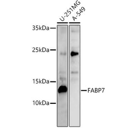
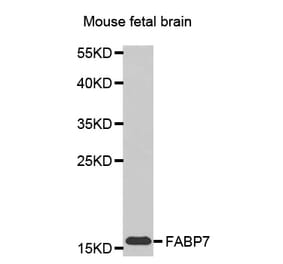
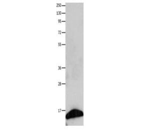
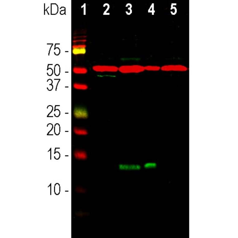
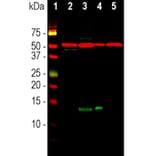
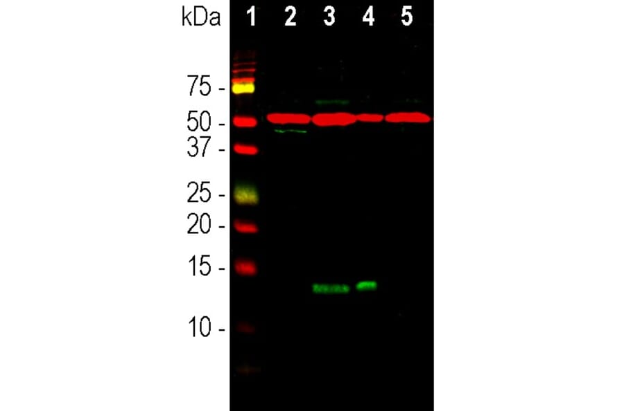
Western blot analysis of whole brain and neuron-glial cell culture lysates using Anti-FABP7 Antibody (A104339), at a dilution of 1:2,000, in green, and Anti-beta Tubulin Antibody [1B12] (A85428), at a dilution of 1:10,000, in red. The lanes contain: [Lane 1] protein standard, [Lane 2] adult rat brain, [Lane 3] E18 embryonic rat brain, [Lane 4] rat neuron-glial cell culture, and [Lane 5] adult mouse brain lysates. The band at ~14kDa corresponds to FABP7 detected only in developing tissue and cells, while the 50kDa band represents the beta Tubulin protein which is present in all preparations.
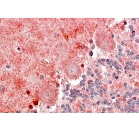
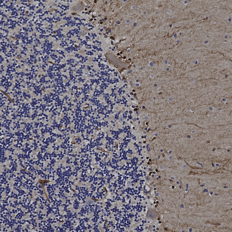
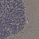
Immunohistochemistry analysis of a formalin fixed paraffin embedded human cerebellum section with Anti-FABP7 Antibody (A104339) at a dilution of 1:2,000. Anti-FABP7 Antibody (A104339) strongly labels the cytoplasm and nuclei of Bergmann glia cells as well as the molecular layer neuropil. Note: this antibody performs well in testing with both 4% PFA and standard NBF fixed rat, mouse and human tissues.
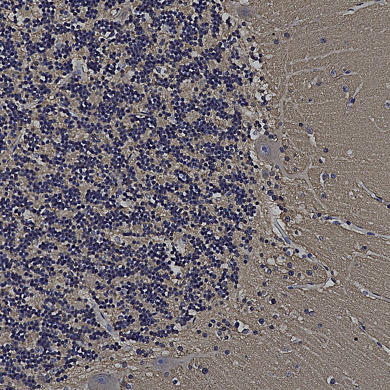
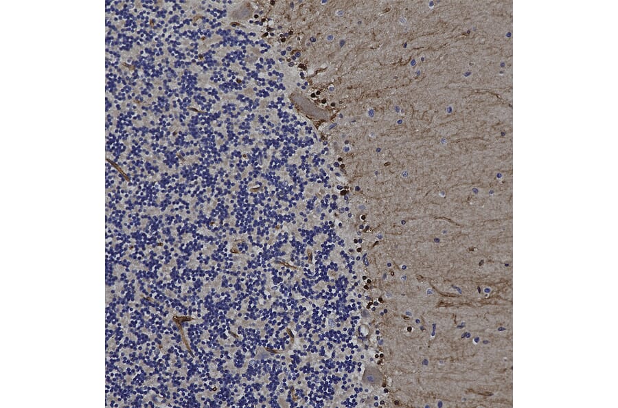
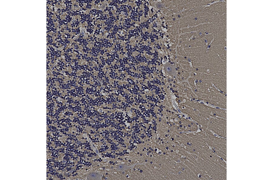
Immunohistochemistry analysis of a formalin fixed paraffin embedded human cerebellum section with Anti-GAP43 Antibody (A85394) at a dilution of 1:2,000 detected with DAB (brown) using the Vector Elite ABC-HRP detection and reagents with citra buffer retrieval. Counterstained with Hematoxylin (blue). The Anti-FABP7 Antibody (A104339) strongly labels cells and neuropil within the molecular layer and processes in the granular layer. Note: this antibody performs well in testing with both 4% PFA and standard NBF fixed rat, mouse and human tissues.
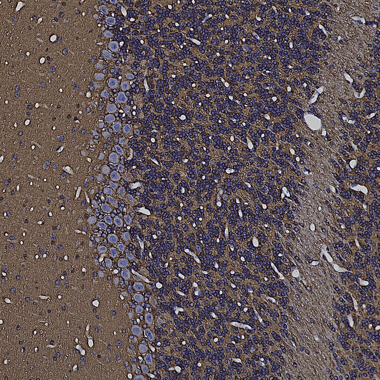
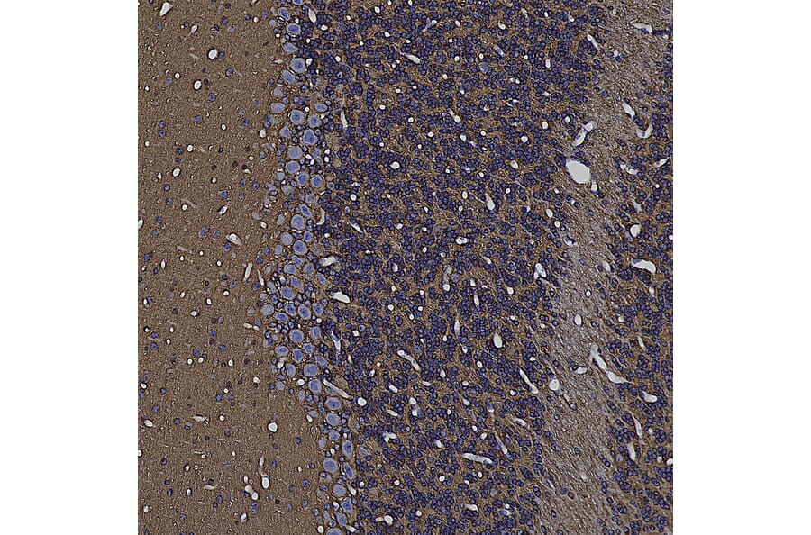
Immunohistochemistry analysis of a formalin fixed paraffin embedded mouse cerebellum section with Anti-GAP43 Antibody (A85394) at a dilution of 1:2,000 detected with DAB (brown) using the Vector Elite ABC-HRP detection and reagents with citra buffer retrieval. Counterstained with Hematoxylin (blue). The Anti-FABP7 Antibody (A104339) strongly labels cells and neuropil within the molecular layer and processes in the granular layer. Note: this antibody performs well in testing with both 4% PFA and standard NBF fixed rat, mouse and human tissues.


![Immunofluorescence - Anti-FABP7 Antibody [2A84] (A270549) - Antibodies.com](https://cdn.antibodies.com/image/catalog/270/A270549_1.jpg?profile=product_alternative)