This polyclonal rabbit anti-FLAG antibody was raised against the FLAG epitope tag and specifically recognizes the DYKDDDDK sequence fused to the N- or C-terminus of recombinant proteins.
CF® Dye and biotin conjugates: Add 0.5 ml dH2O for reconstitution. HRP or DNP conjugates: Add 1 ml dH2O for reconstitution.
Formulation
Supplied in Phosphate Buffered Saline containing 50% glycerol, 2 mg/ml BSA, and 0.05% Sodium Azide.
Storage
Shipped at +4°C. Upon delivery aliquot and store at -20°C. Avoid freeze/thaw cycles. This product is also photosensitive and should be protected from light. Should this product contain a precipitate we recommend microcentrifugation before use. CF® Dyes are guaranteed for at least 6 months from data of receipt when stored correctly.
General Notes
Looking for a specific protein conjugate to simplify your workflow? We offer a library of over 2,000 targets conjugated to your choice of CF® dye. To enquire about a custom product, contact us directly.
Synonyms
DDDDK epitope tag, DDDDK tag, DDDK, ddk, DYKDDDDK, DYKDDDDK epitope tag, DYKDDDDK tag, ECS epitope tag, ECS tag, Enterokinase Cleavage Site epitope tag, Enterokinase Cleavage Site tag, FLAG
Disclaimer
This product is for research use only. It is not intended for diagnostic or therapeutic use.
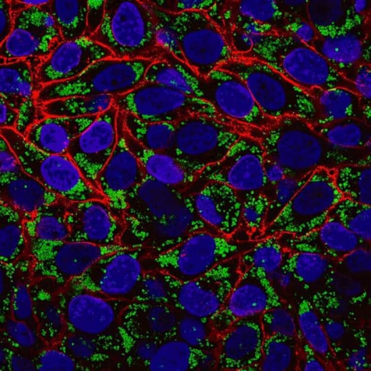
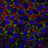
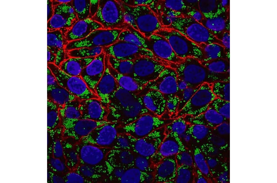
Fixed oleic acid–treated HeLa cells stained with LipidSpot™ 488 to visualize lipid droplets (green), CF®594 wheat germ agglutinin to label the cell surface (red), and Hoechst 33258 to stain nuclei (blue).
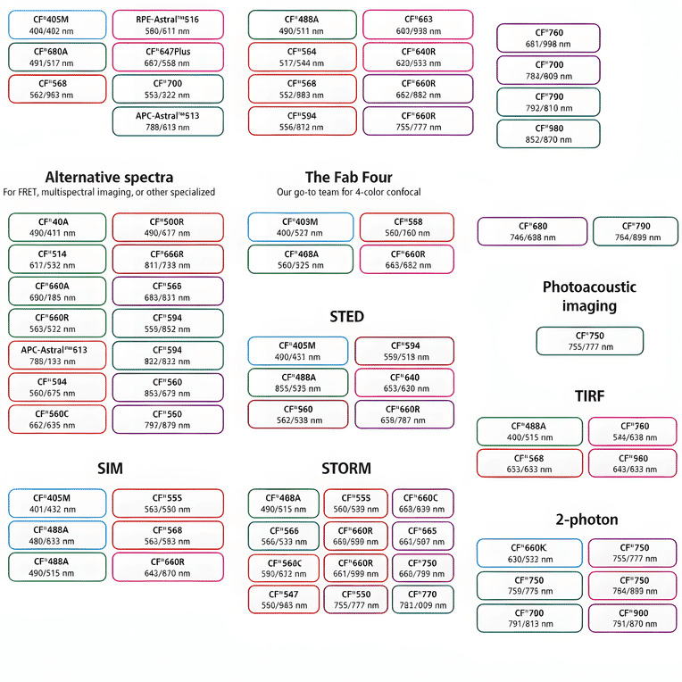
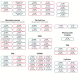
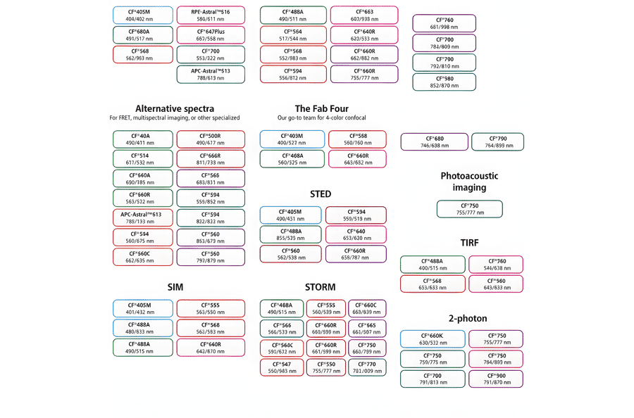
This chart summarizes commonly used CF® dyes grouped by their suitability for specific imaging modalities, including alternative spectra applications, four-color confocal imaging, near-infrared western blotting, photoacoustic imaging, STED, SIM, STORM, TIRF, and two-photon microscopy. Each dye is shown with its characteristic excitation and emission wavelengths (nm), providing a practical reference for selecting spectrally compatible dyes and optimizing multicolor experimental design across a range of fluorescence techniques.
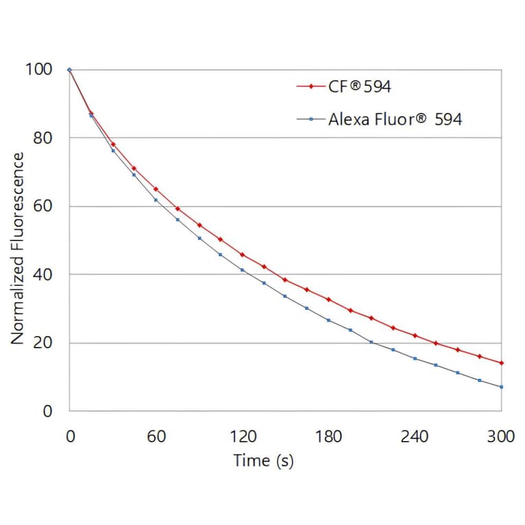
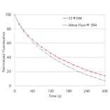
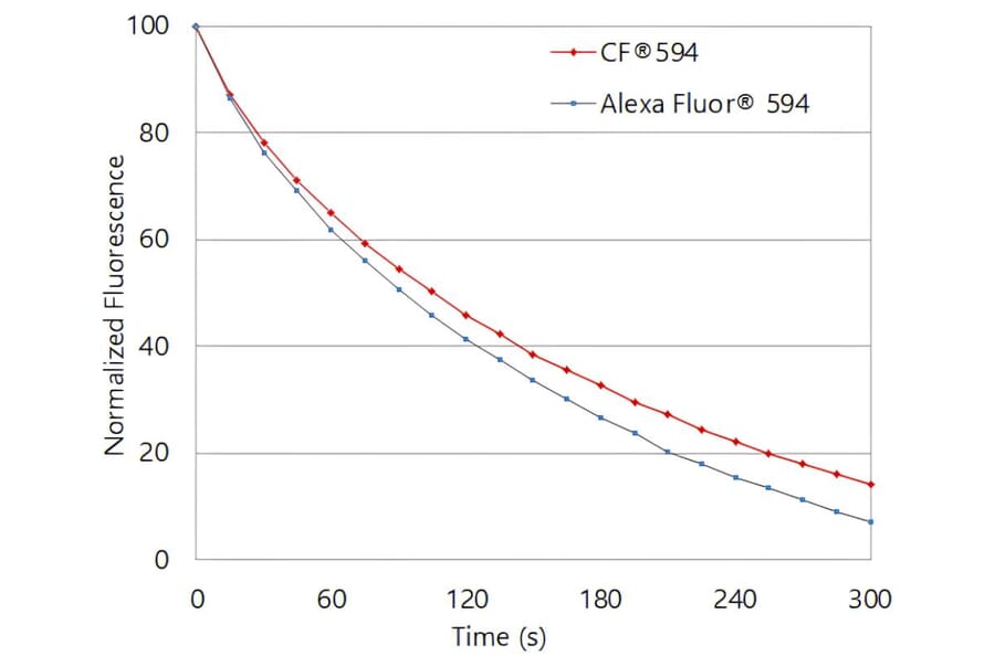
Photostability comparison of CF®594 and Alexa Fluor® 594. Jurkat cells were stained with mouse anti-CD3 primary antibody followed by CF®594 or Alexa Fluor® 594 goat anti-mouse IgG secondary antibodies. Cells were continuously illuminated, and fluorescence intensity was monitored over time.
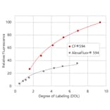
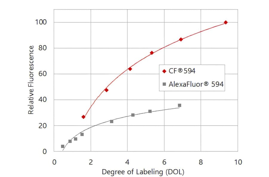
CF®594 produces brighter antibody conjugates than Alexa Fluor® 594. Relative fluorescence intensity of goat anti-mouse IgG conjugates is shown as a function of degree of labeling (DOL, dye molecules per antibody).
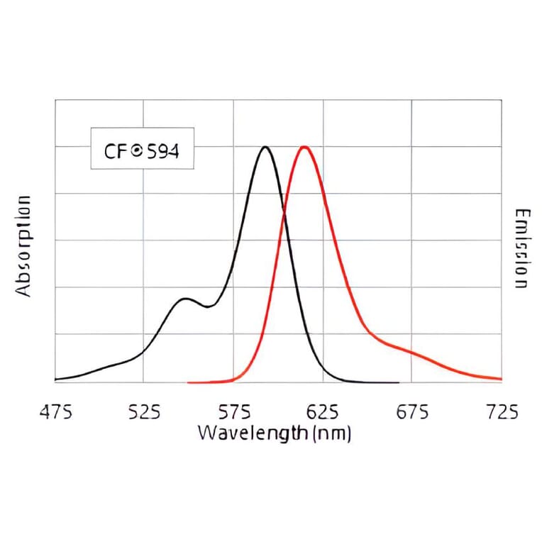
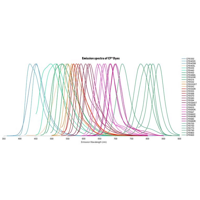
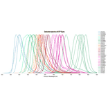
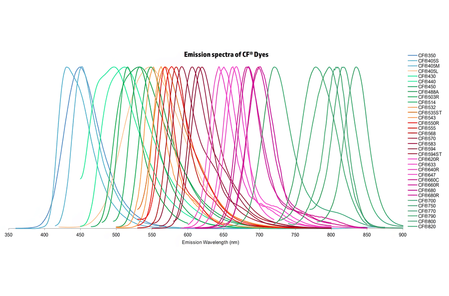
Normalized emission spectra of the CF® dye family spanning the visible to near-infrared range are shown, illustrating the spectral diversity and overlap between dyes. Curves represent relative fluorescence intensity as a function of emission wavelength (nm), with peak positions corresponding to each dye’s characteristic emission maximum. This reference highlights the broad coverage of CF® dyes for multicolor fluorescence applications and aids in selecting compatible dye combinations for imaging, flow cytometry, and other fluorescence-based assays.
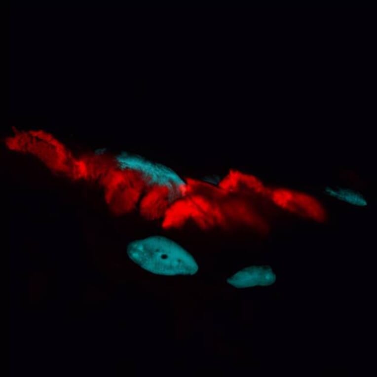
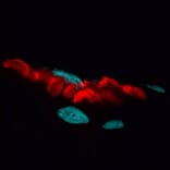
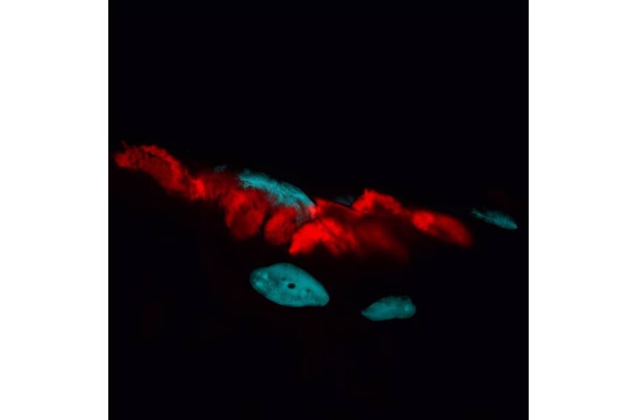
Neuromuscular junctions in a rat skeletal muscle section stained with CF®594 a-bungarotoxin to label acetylcholine receptors (red), with nuclei counterstained using DAPI (blue).
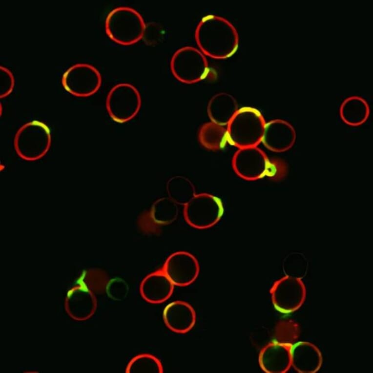
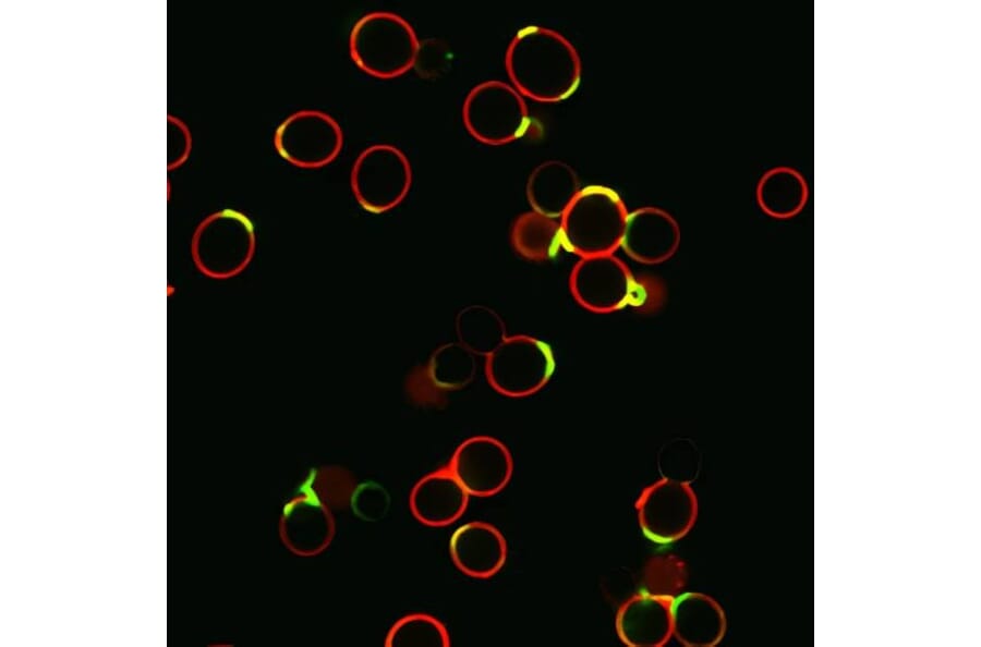
Saccharomyces cerevisiae yeast stained with CF®594 wheat germ agglutinin to label cell walls (red) and CF®488A concanavalin A to label bud scars (green).