Mouse monoclonal [TU-30] antibody to gamma Tubulin.
Specificity
This antibody recognizes the C terminus of human gamma Tubulin, a 48 kDa structural constituent of cytoskeleton and microtubule organizing center (MTOC). The epitope is located in the amino acid sequence TRPDYI (a.a. 439-444 in human), which is present on human gamma Tubulin 1 but not on human gamma Tubulin 2.
Applications
ICC, WB, Flow Cytometry (Intracellular)
Dilutions
IHC: 1-2 µg/ml. Staining technique: (a) Fix cells for 10 min in methanol at -20°C and for 6 min in acetone at -20°C; (b) Fix cells directly in methanol for 10 min at -20°C or in acetone for 10 min at -20°C. Positive Control: P-19 murine embryonal carcinoma cell line, 3T3 murine fibroblasts. The antibody TU-30 stains only fixed cells, WB: Recommended dilution 1-2 µg/ml; Reducing conditions.
Reactivity
Protozoa, Chicken, Bovine, Rat, Mouse, Porcine, Human, Plants
Immunogen
C-terminal peptide of gamma-tubulin counjugated to KLH.
Host
Mouse
Clonality
Monoclonal
Clone ID
TU-30
Isotype
IgG1
Conjugate
Unconjugated
Purification
Protein A chromatography.
Concentration
1 mg/ml
Predicted MW
48 kDa
Product Form
Liquid
Formulation
Supplied in Phosphate Buffered Saline, pH 7.4, with 15 mM Sodium Azide.
Storage
Shipped at 4°C. Upon delivery aliquot and store at -20°C. Avoid freeze/thaw cycles.
Immunocytochemistry staining of P19X1 mouse embryonal carcinoma cell line using Anti-gamma Tubulin Antibody [TU-30] (A86254), (detection by secondary antibody Goat anti-mouse Cy3). Nuclei were stained with DAPI (blue).
Immunocytochemistry staining of murine fibroblasts using Anti-gamma Tubulin Antibody [TU-30] (A86254) direct conjugate with Dyomics 547, red). Nuclei were stained with DAPI (blue).
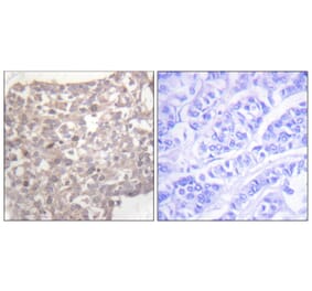
Immunocytochemistry staining of microtubular networks in 3T3 mouse fibroblasts.A - metaphase; B - anaphase; C - telophaseGamma-tubulin (red) stained using Anti-gamma Tubulin Antibody [TU-30] (A86254) , alpha-tubulin (green) with polyclonal anti-alpha-tubulin antibody and nuclei with DAPI (blue).
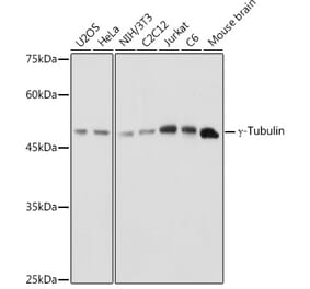
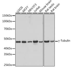
Western blotting analysis of human gamma-tubulin using Anti-gamma Tubulin Antibody [TU-30] (A86254) on lysates of various cell lines under reducing and non-reducing conditions. Nitrocellulose membrane was probed with 2 µg/ml of mouse anti-gamma-tubulin monoclonal antibody followed by IRDye800-conjugated anti-mouse secondary antibody. A specific band was detected for gamma-tubulin at approximately 46 kDa.
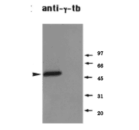
Western blotting analysis of differential reactivity of monoclonal antibodies to ?-tubulin with human ?-tubulin isotypes. (A) Immunoblots of total cell lysates from SH-SY5Y cells, expressing TagRFP-tagged human ?-tubulin 1 (?-Tb1) or ?-tubulin 2 (?-Tb2), probed with ?-tubulin antibodies: Anti-gamma Tubulin Antibody [TU-30] (A86254) and Anti-gamma Tubulin Antibody [TU-32] (A86251), TagRFP (RFP) and GAPDH. In control samples, only secondary anti-mouse Ab was applied. (B) Immunoblots of immobilized GST-tagged human C-terminal regions (a.a. 362-451) of ?-Tb1 or ?-Tb2 probed with Abs to ?-tubulin (TU-30, TU-32) and GST. In control samples, only secondary anti-mouse Ab was applied.


![ICC - Anti-gamma Tubulin Antibody [TU-30] (A86254)](https://cdn.antibodies.com/image/catalog/86/A86254_1.jpg?profile=product_top)
![Flow Cytometry - Anti-gamma Tubulin Antibody [TU-30] (A86254)](https://cdn.antibodies.com/image/catalog/86/A86254_2.jpg?profile=product_top)
![ICC - Anti-gamma Tubulin Antibody [TU-30] (A86254)](https://cdn.antibodies.com/image/catalog/86/A86254_3.jpg?profile=product_top)
![ICC - Anti-gamma Tubulin Antibody [TU-30] (A86254)](https://cdn.antibodies.com/image/catalog/86/A86254_4.jpg?profile=product_top)
![WB - Anti-gamma Tubulin Antibody [TU-30] (A86254)](https://cdn.antibodies.com/image/catalog/86/A86254_5.jpg?profile=product_top)
![WB - Anti-gamma Tubulin Antibody [TU-30] (A86254)](https://cdn.antibodies.com/image/catalog/86/A86254_6.jpg?profile=product_top)
![ICC - Anti-gamma Tubulin Antibody [TU-30] (A86254)](https://cdn.antibodies.com/image/catalog/86/A86254_1.jpg?profile=product_top_thumb)
![Flow Cytometry - Anti-gamma Tubulin Antibody [TU-30] (A86254)](https://cdn.antibodies.com/image/catalog/86/A86254_2.jpg?profile=product_top_thumb)
![ICC - Anti-gamma Tubulin Antibody [TU-30] (A86254)](https://cdn.antibodies.com/image/catalog/86/A86254_3.jpg?profile=product_top_thumb)
![ICC - Anti-gamma Tubulin Antibody [TU-30] (A86254)](https://cdn.antibodies.com/image/catalog/86/A86254_4.jpg?profile=product_top_thumb)
![WB - Anti-gamma Tubulin Antibody [TU-30] (A86254)](https://cdn.antibodies.com/image/catalog/86/A86254_5.jpg?profile=product_top_thumb)
![WB - Anti-gamma Tubulin Antibody [TU-30] (A86254)](https://cdn.antibodies.com/image/catalog/86/A86254_6.jpg?profile=product_top_thumb)
![ICC - Anti-gamma Tubulin Antibody [TU-30] (A86254)](https://cdn.antibodies.com/image/catalog/86/A86254_1.jpg?profile=product_image)
![Flow Cytometry - Anti-gamma Tubulin Antibody [TU-30] (A86254)](https://cdn.antibodies.com/image/catalog/86/A86254_2.jpg?profile=product_image)
![ICC - Anti-gamma Tubulin Antibody [TU-30] (A86254)](https://cdn.antibodies.com/image/catalog/86/A86254_3.jpg?profile=product_image)
![ICC - Anti-gamma Tubulin Antibody [TU-30] (A86254)](https://cdn.antibodies.com/image/catalog/86/A86254_4.jpg?profile=product_image)
![WB - Anti-gamma Tubulin Antibody [TU-30] (A86254)](https://cdn.antibodies.com/image/catalog/86/A86254_5.jpg?profile=product_image)
![WB - Anti-gamma Tubulin Antibody [TU-30] (A86254)](https://cdn.antibodies.com/image/catalog/86/A86254_6.jpg?profile=product_image)
![WB - Anti-gamma Tubulin Antibody [TU-32] (A86251)](https://cdn.antibodies.com/image/catalog/86/A86251_1.jpg?profile=product_alternative)