This antibody recognises GATA3 also known as trans-acting T-cell-specific transcription factor GATA-3 or GATA-binding factor 3.The GATA3 protein is a member of the GATA family of transcription factors which has received its name from binding DNA sequences containing the GATA consensus motif 5'-[AT]GATA[AG]-3' (P15976). GATA3 has been shown to play a critical role in T cell development and the differentiation of T helper 2 (Th2) cells. In addition, GATA3 has been implied to have tissue specific functions such as a regulatory role in the development of the mammary gland epithelium and skin tissue. Aberrant GATA3 levels have also been identified in a variety of cancers including breast cancer. Indeed, GATA3 has become a marker for certain breast cancer types and GATA3 antibodies are commonly used for the immunohistochemical evaluation of breast cancer samples.
Purified partial recombinant recombinant human GATA3 protein expressed in E. coli.
Host
Mouse
Clonality
Monoclonal
Clone ID
1A12-1D9
Isotype
IgG1
Conjugate
Unconjugated
Purification
Protein A affinity chromatography of tissue culture supernatant.
Concentration
1 mg/ml
Molecular Weight
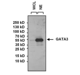
Approximately 55 kDa in SH-SY5Y cell lysate and SH-SY5Y nuclear extract.
Product Form
Liquid
Formulation
Supplied in Phosphate Buffered Saline with 1 mg/ml BSA, 30% Glycerol, and <0.1% Sodium Azide.
Storage
Shipped at ambient temperature. Upon delivery aliquot and store at -20°C. When thawed, aliquot the sample as needed. Short term (up to 4 weeks): store at 4°C. Long term: store at -20°C. Avoid freeze / thaw cycles. Storage in frost free freezers is not recommended.
General Notes
Mouse anti GATA3 antibody clone 1A12-1D9 recognizes GATA3 also known as trans-acting T-cell-specific transcription factor GATA-3 or GATA-binding factor 3.The GATA3 protein is a member of the GATA family of transcription factors which has received its name from binding DNA sequences containing the GATA consensus motif 5'-[AT]GATA[AG]-3' (P15976). GATA3 has been shown to play a critical role in T cell development and the differentiation of T helper 2 (Th2) cells (Ho et al. 1991). In addition, GATA3 has been implied to have tissue specific functions such as a regulatory role in the development of the mammary gland epithelium and skin tissue (Chou et al. 2010).Aberrant GATA3 levels have also been identified in a variety of cancers including breast cancer. Indeed, GATA3 has become a marker for certain breast cancer types and GATA3 antibodies are commonly used for the immunohistochemical evaluation of breast cancer samples (Krings et al. 2014).
Western Blot - Anti-GATA3 Antibody [1A12-1D9] (A279065)
Western Blot analysis of GATA3 was performed by loading 15ug of SH-SY5Y whole cell lysate (WCL) and SH-SY5Y nuclear extract (NE) onto a 4-20% Tris-HCl polyacrylamide gel. Proteins were transferred to a PVDF membrane and blocked for at least 1 hour. The membrane was probed with Anti-GATA3 Antibody [1A12-1D9] (A279065) at a dilution of 1/1000 overnight at 4°C on a rocking platform, washed in TBS-0.1%Tween-20, and probed with an HRP-conjugated goat anti-mouse IgG secondary antibody for 1 hour. Chemiluminescent detection was performed.
Immunofluorescence analysis of GATA3 (green) in MCF7 cells. Formalin-fixed cells were permeabilized with 0.1% Triton X-100 in TBS for 10 minutes at room temperature. Cells were blocked with 1% Blocker BSA for 15 minutes at room temperature. Cells were probed without (left panel) or with (right panel) Anti-GATA3 Antibody [1A12-1D9] (A279065) at a dilution of 1/50 for at least 1 hour at room temperature, washed with PBS, and incubated with a DyLight 488-conjugated goat anti-mouse IgG secondary antibody for 30 minutes at room temperature. F-Actin (red) was stained with phalloidin (red) and nuclei (blue) were stained with Hoechst 33342 dye. Images were taken at 20x magnification.
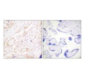
Immunohistochemistry analysis of GATA3 showing staining in the nucleus of paraffin-embedded human breast carcinoma (right) compared to a negative control without primary antibody (left). To expose target proteins, antigen retrieval was performed using 10mM sodium citrate (pH 6.0), microwaved for 8-15 min. Following antigen retrieval, tissues were blocked in 3% H2O2-methanol for 15 min at room temperature, washed with dH2O and PBS, and then probed with Anti-GATA3 Antibody [1A12-1D9] (A279065) diluted in 3% BSA-PBS at a dilution of 1/1000 overnight at 4°C in a humidified chamber. Tissues were washed extensively in PBST and detection was performed using an HRP-conjugated secondary antibody followed by colorimetric detection using a DAB kit. Tissues were counterstained with hematoxylin and dehydrated with ethanol and xylene to prep for mounting.
Publishing research using Anti-GATA3 Antibody [1A12-1D9] (A279065)? Please let us know so that we can list the citation on this page.
Alternative products to Anti-GATA3 Antibody [1A12-1D9] (A279065)


![Western Blot - Anti-GATA3 Antibody [1A12-1D9] (A279065) - Antibodies.com](https://cdn.antibodies.com/image/catalog/279/A279065_1.jpg?profile=product_top)
![Immunofluorescence - Anti-GATA3 Antibody [1A12-1D9] (A279065) - Antibodies.com](https://cdn.antibodies.com/image/catalog/279/A279065_2.jpg?profile=product_top)
![Immunohistochemistry - Anti-GATA3 Antibody [1A12-1D9] (A279065) - Antibodies.com](https://cdn.antibodies.com/image/catalog/279/A279065_3.jpg?profile=product_top)
![Western Blot - Anti-GATA3 Antibody [1A12-1D9] (A279065) - Antibodies.com](https://cdn.antibodies.com/image/catalog/279/A279065_1.jpg?profile=product_top_thumb)
![Immunofluorescence - Anti-GATA3 Antibody [1A12-1D9] (A279065) - Antibodies.com](https://cdn.antibodies.com/image/catalog/279/A279065_2.jpg?profile=product_top_thumb)
![Immunohistochemistry - Anti-GATA3 Antibody [1A12-1D9] (A279065) - Antibodies.com](https://cdn.antibodies.com/image/catalog/279/A279065_3.jpg?profile=product_top_thumb)
![Western Blot - Anti-GATA3 Antibody [1A12-1D9] (A279065) - Antibodies.com](https://cdn.antibodies.com/image/catalog/279/A279065_1.jpg?profile=product_image)
![Immunofluorescence - Anti-GATA3 Antibody [1A12-1D9] (A279065) - Antibodies.com](https://cdn.antibodies.com/image/catalog/279/A279065_2.jpg?profile=product_image)
![Immunohistochemistry - Anti-GATA3 Antibody [1A12-1D9] (A279065) - Antibodies.com](https://cdn.antibodies.com/image/catalog/279/A279065_3.jpg?profile=product_image)
![Immunohistochemistry - Anti-GATA3 Antibody [GATA3/6664] (A277623) - Antibodies.com](https://cdn.antibodies.com/image/catalog/277/A277623_1.jpg?profile=product_alternative)
![Immunohistochemistry - Anti-GATA3 Antibody [GATA3/6664] - BSA and Azide free (A278211) - Antibodies.com](https://cdn.antibodies.com/image/catalog/278/A278211_1.jpg?profile=product_alternative)

![Protein Array - Anti-GATA3 Antibody [GATA3/2445] (A251878) - Antibodies.com](https://cdn.antibodies.com/image/catalog/248/A248697_1.jpg?profile=product_alternative)
![Protein Array - Anti-GATA3 Antibody [GATA3/2445] - BSA and Azide free (A248697) - Antibodies.com](https://cdn.antibodies.com/image/catalog/251/A251879_1.jpg?profile=product_alternative)