

Unconjugated
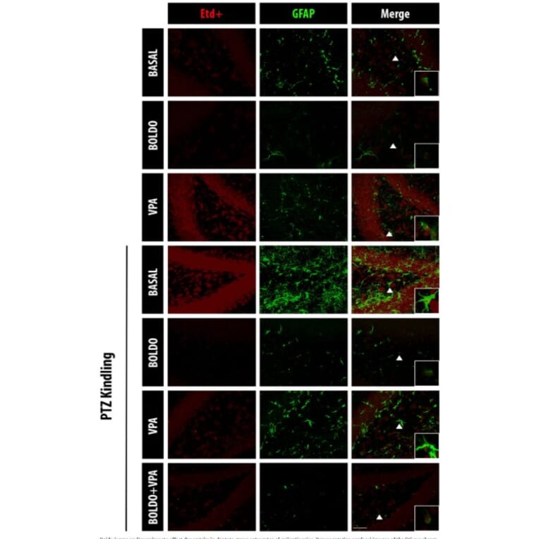
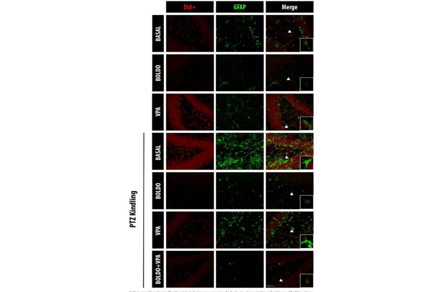
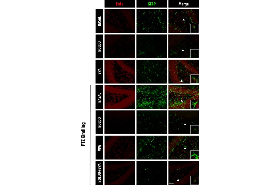
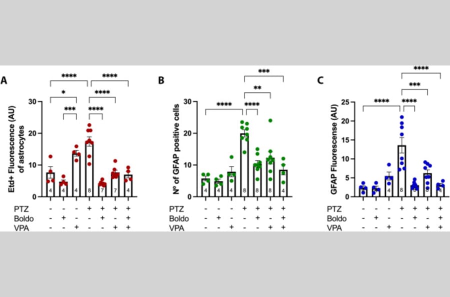
Epilepsy is a chronic neurological disorder characterized by a propensity for seizures due to an imbalance between excitatory and inhibitory brain activity. This condition also induces neuroinflammation, which contributes to disease progression. Given that hemichannels (HCs) permeabilize the cell membrane of glia playing a critical role in neuroinflammation, we investigated the antiepileptic potential of Boldo (Peumus boldus M.), an endemic Chilean tree containing several bioactive molecules including boldine, a HC inhibitor. Mice were treated with pulverized Boldo leaves, the antiseizure medication valproate, or a combination of both for 5 days. Seizure severity was assessed in a pentylenetetrazole-induced kindling mouse model. Using the dye uptake technique, we evaluated the membrane permeability in hippocampal astrocytes, microglia, and neurons. Additionally, we analyzed astroglial and microglial reactivity and measured levels of pro-inflammatory cytokines (IL-1ß, IL6, and TNF-a). Both Boldo and valproate significantly reduced seizure severity. However, distinct mechanisms were observed. Valproate administration increased dye uptake in control animals and enhanced glial reactivity, corroborating its established ability to stimulate hemichannel activity. Conversely, Boldo treatment, either alone or in conjunction with valproate, reduced these parameters, consistent with its HC-blocking properties. Importantly, Boldo was more effective than valproate in reducing plasmatic levels of inflammatory and oxidative stress markers. These findings indicate that Boldo, by inhibiting these HCs, could provide a valuable therapeutic strategy to mitigate neuroinflammation in epilepsy, highlighting the clinical potential of this readily available medicinal herb.
Toxoplasma (T.) gondii is an obligate intracellular parasite with a worldwide distribution. Congenital infection can lead to severe pathological alterations in the brain. To examine the effects of toxoplasmosis in the fetal brain, pregnant guinea pigs are infected with T. gondii oocysts on gestation day 23 and dissected 10, 17 and 25 days afterwards. We show the neocortex to represent a target region of T. gondii and the parasite to infect neural progenitor cells (NPCs), neurons and astrocytes in the fetal brain. Importantly, we observe a significant reduction in neuron number at end-neurogenesis and find a marked reduction in NPC count, indicating that impaired neurogenesis underlies the neuronal decrease in infected fetuses. Moreover, we observe focal microglioses to be associated with T. gondii in the fetal brain. Our findings expand the understanding of the pathophysiology of congenital toxoplasmosis, especially contributing to the development of cortical malformations.
Lucy Xu, Harvard Medical School

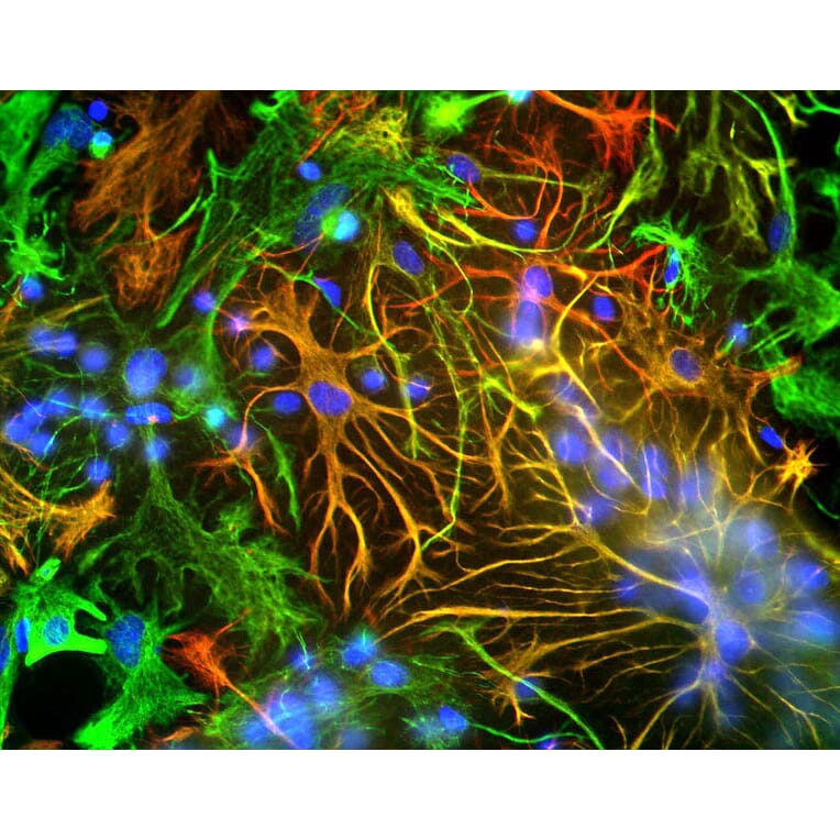

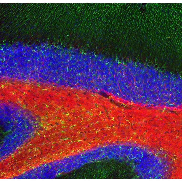


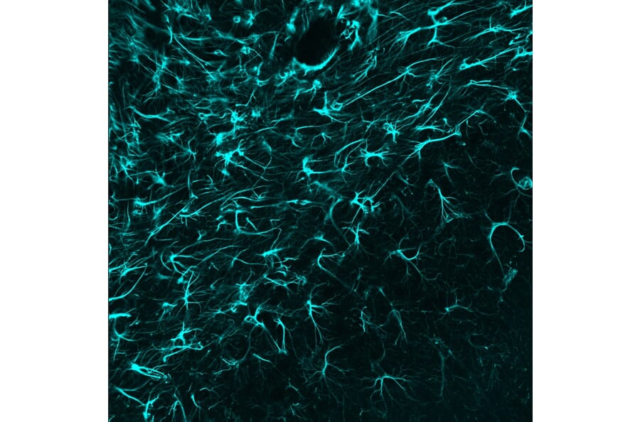


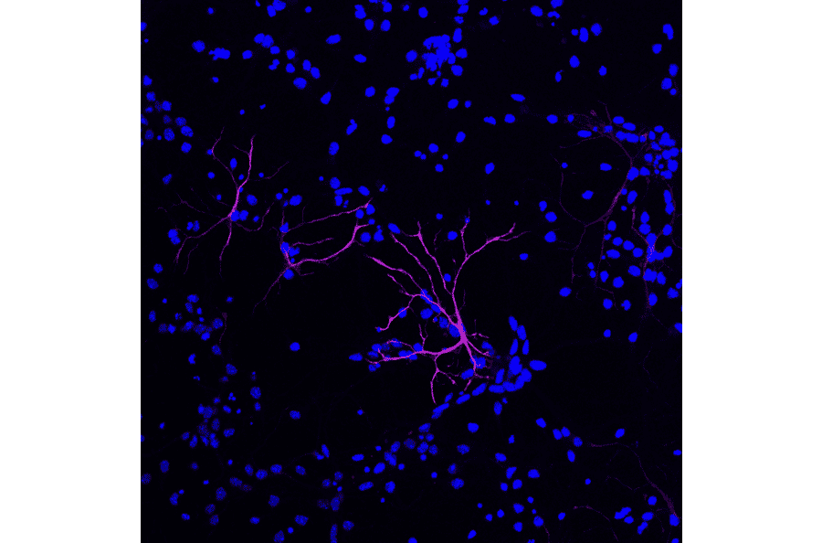
![Immunofluorescence - Anti-GFAP Antibody [2A5] (A104314) - Antibodies.com](https://cdn.antibodies.com/image/catalog/104/A104314_1.jpg?profile=product_alternative)
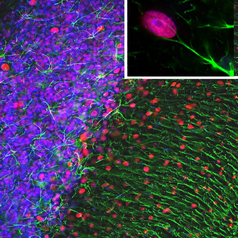
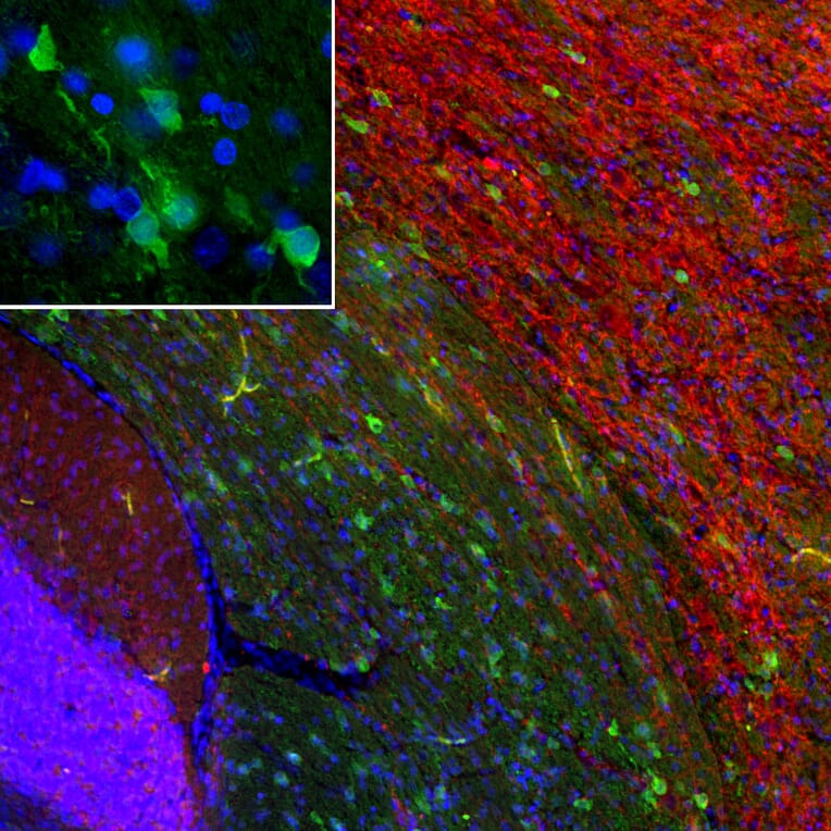
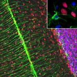
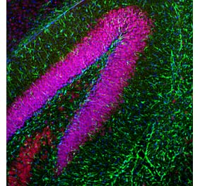
![Immunofluorescence - Anti-GFAP Antibody [5C10] (A85422) - Antibodies.com](https://cdn.antibodies.com/image/catalog/85/A85422_1.jpg?profile=product_alternative)
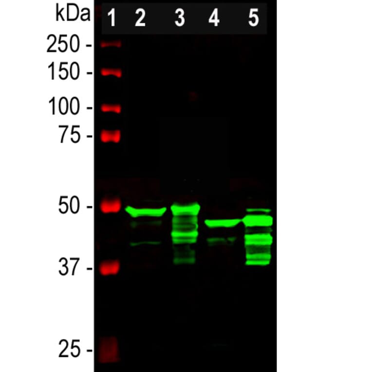
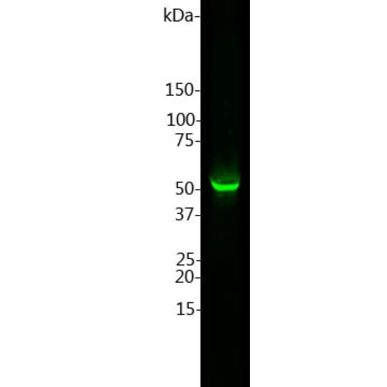
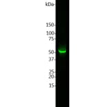
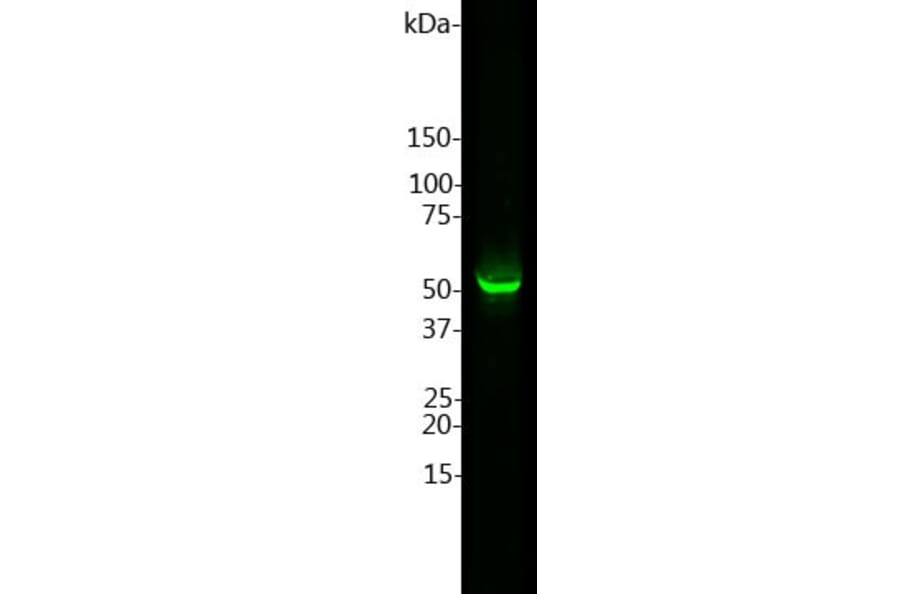
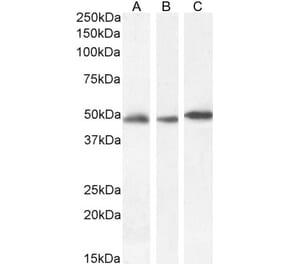
![Western Blot - Anti-GFAP Antibody [GA-5] - BSA and Azide free (A251887) - Antibodies.com](https://cdn.antibodies.com/image/catalog/251/A251887_1.jpg?profile=product_alternative)
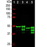
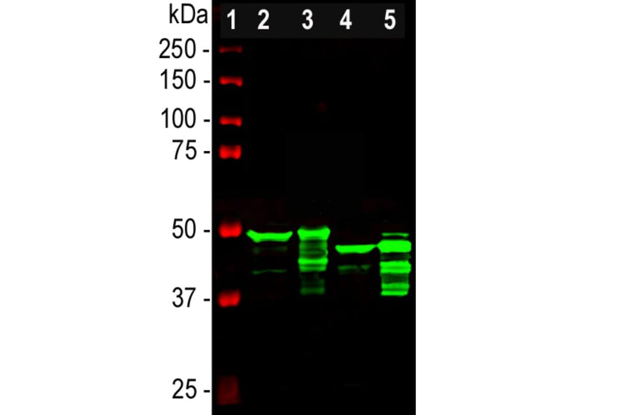
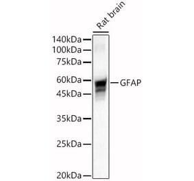
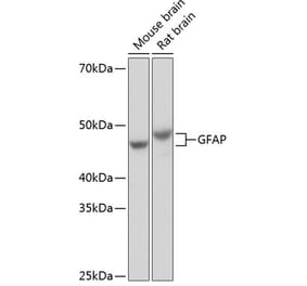
![Western Blot - Anti-GFAP Antibody [GA-5] (A248705) - Antibodies.com](https://cdn.antibodies.com/image/catalog/248/A248705_1.jpg?profile=product_alternative)
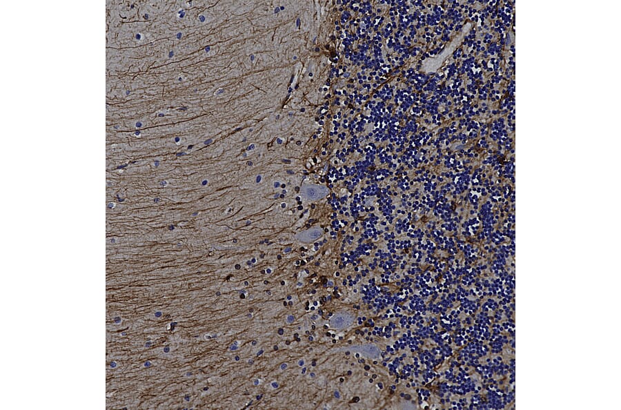
![Immunohistochemistry - Anti-GFAP Antibody [SPM507] - BSA and Azide free (A248707) - Antibodies.com](https://cdn.antibodies.com/image/catalog/251/A251889_1.jpg?profile=product_alternative)
![Immunohistochemistry - Anti-GFAP Antibody [SPM248] - BSA and Azide free (A248706) - Antibodies.com](https://cdn.antibodies.com/image/catalog/251/A251888_1.jpg?profile=product_alternative)
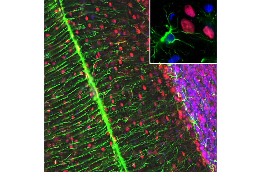
![Immunohistochemistry - Anti-GFAP Antibody [GA-5 + ASTRO/789] (A251890) - Antibodies.com](https://cdn.antibodies.com/image/catalog/248/A248709_1.jpg?profile=product_alternative)
![Immunohistochemistry - Anti-GFAP Antibody [GA-5 + ASTRO/789] - BSA and Azide free (A248709) - Antibodies.com](https://cdn.antibodies.com/image/catalog/251/A251891_1.jpg?profile=product_alternative)
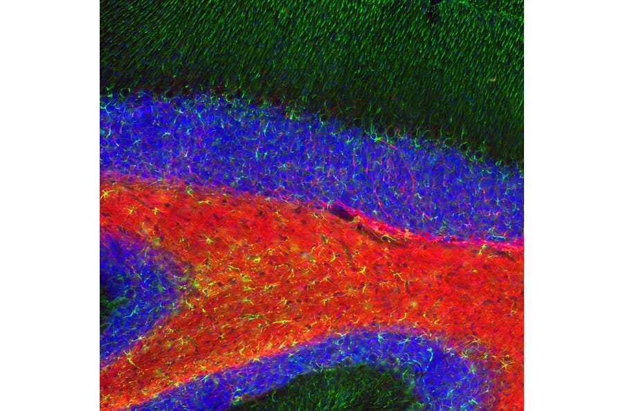
![Immunohistochemistry - Anti-GFAP Antibody [SPM248] (A251891) - Antibodies.com](https://cdn.antibodies.com/image/catalog/248/A248706_1.jpg?profile=product_alternative)
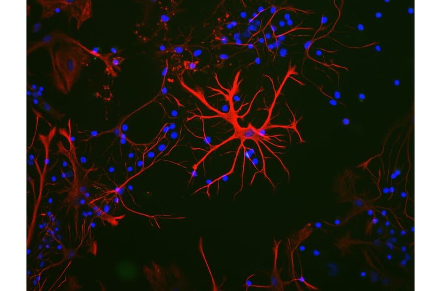
![Immunohistochemistry - Anti-GFAP Antibody [rASTRO/789] - BSA and Azide free (A248711) - Antibodies.com](https://cdn.antibodies.com/image/catalog/251/A251893_1.jpg?profile=product_alternative)