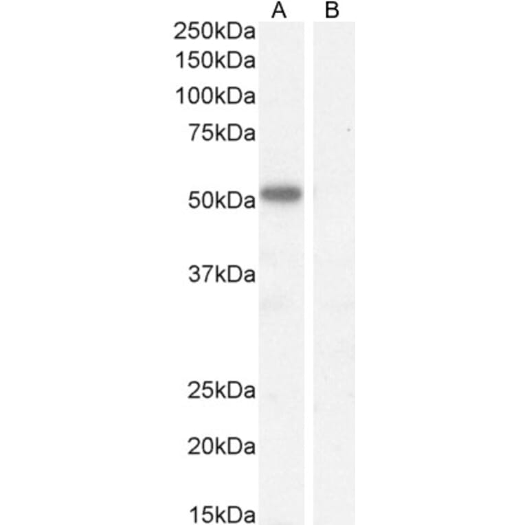
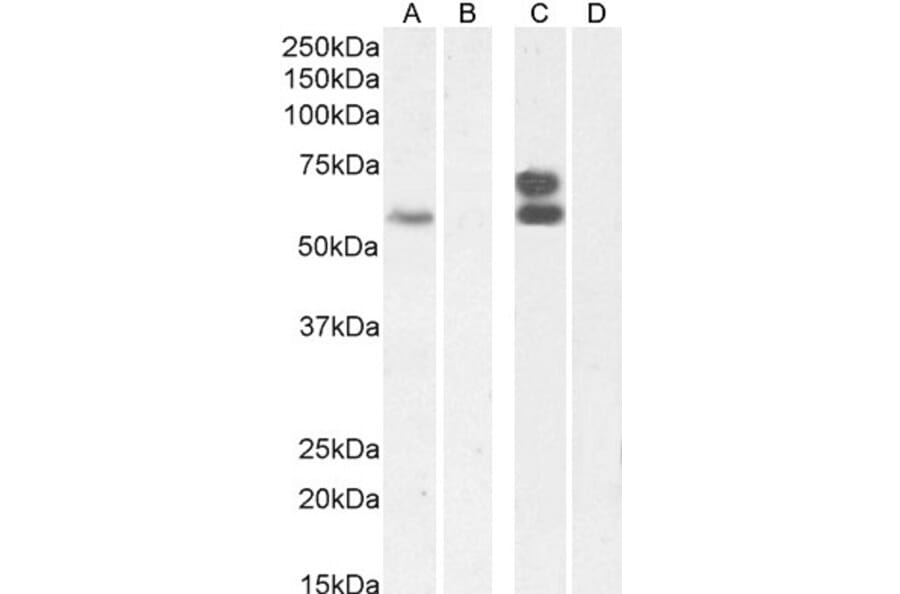
IKZF4 expression in Mouse Heart lysate analyzed by western blot. Cells were lysed in RIPA buffer and 35µg protein was run per lane. Primary antibody incubation was performed with Anti-IKZF4 Antibody (A326255) at 1µg/ml and detected by chemiluminescence.
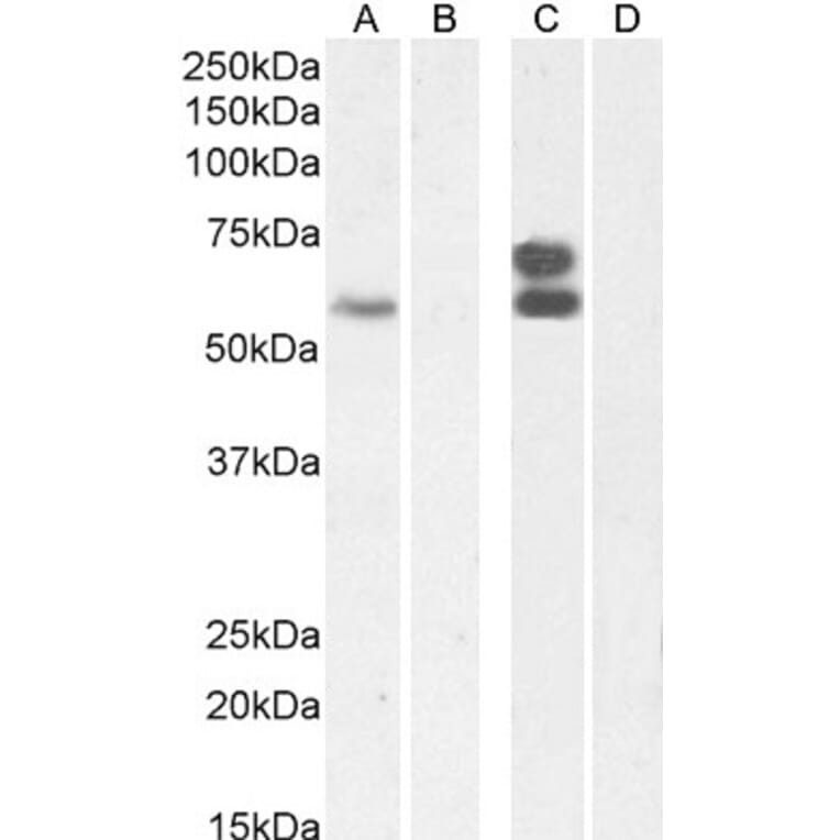
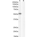
IKZF4 expression in Jurkat nuclear, K562 nuclear lysate (C) + Peptide (D), analyzed by western blot. Cells were lysed in RIPA buffer and 35µg protein was run per lane. Primary antibody incubation was performed with Anti-IKZF4 Antibody (A326255) at 2µg/ml and detected by chemiluminescence.
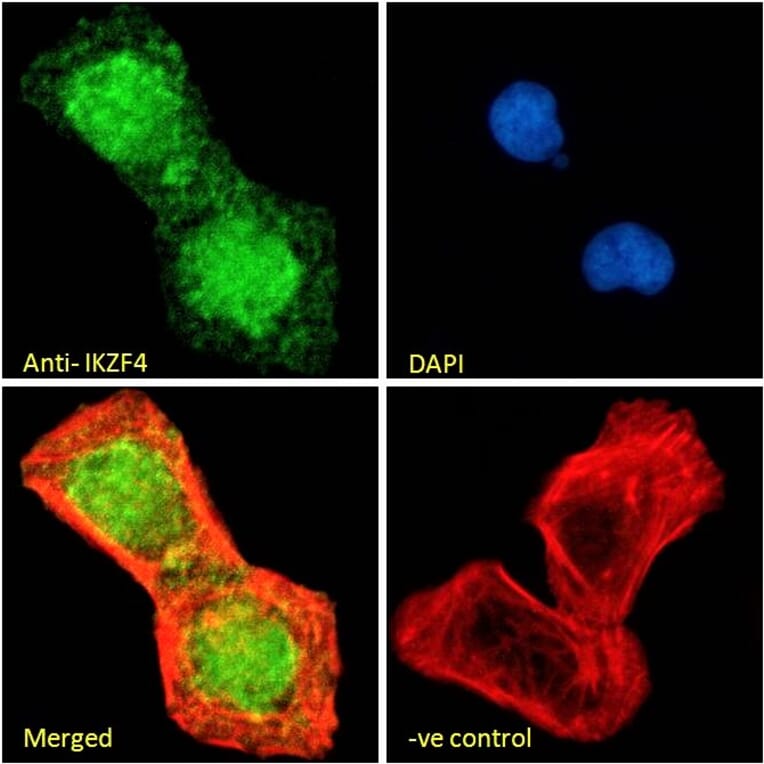
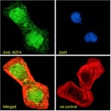
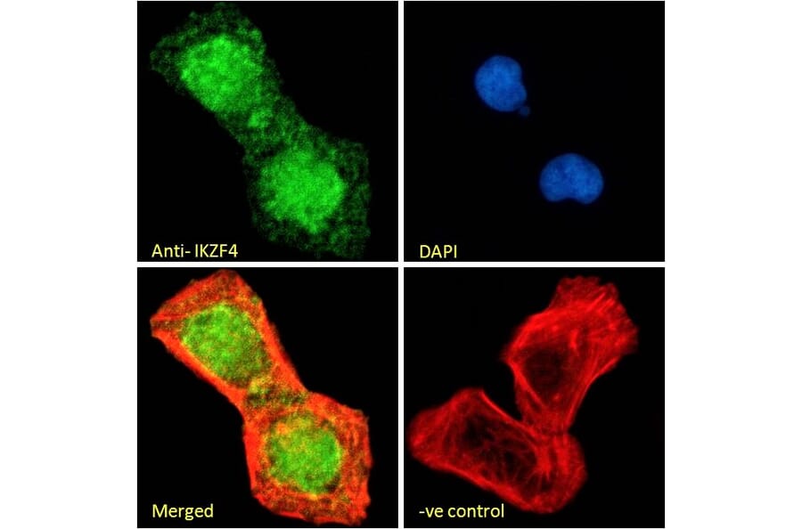
IKZF4 expression in U2OS cells analyzed by immunofluorescence. Cells were permeabilized with 0.15% Triton. Staining was performed with Anti-IKZF4 Antibody (A326255) at 10µg/ml for 1 hour and Alexa Fluor 488 secondary antibody at 2µg/ml. Nuclear staining shown and nuclei were stained with DAPI (blue) while actin filaments were stained with phalloidin (red). Negative control: Goat IgG Isotype Control at 10µg/ml followed by Alexa Fluor 488 secondary antibody at 2µg/ml.
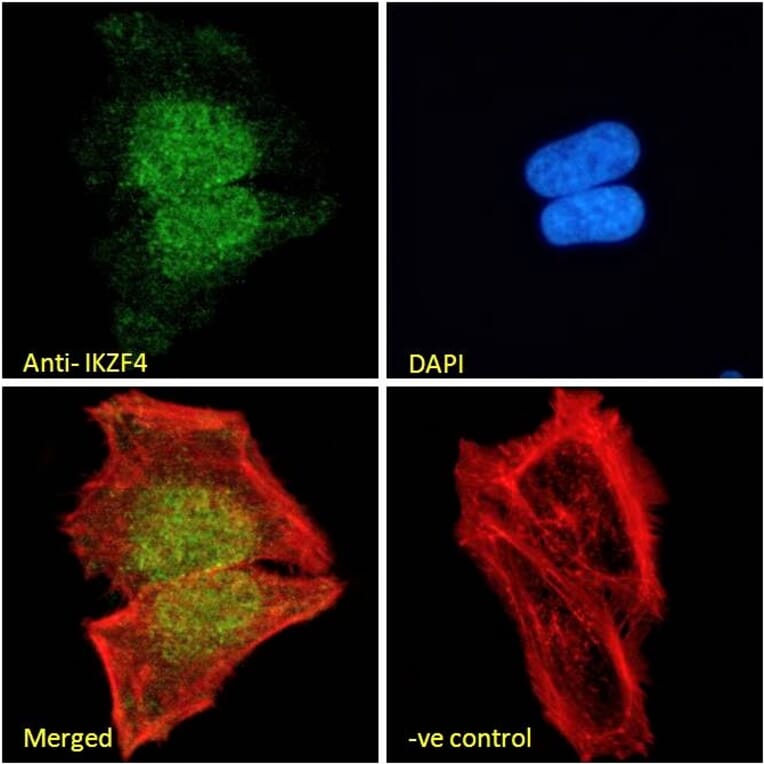
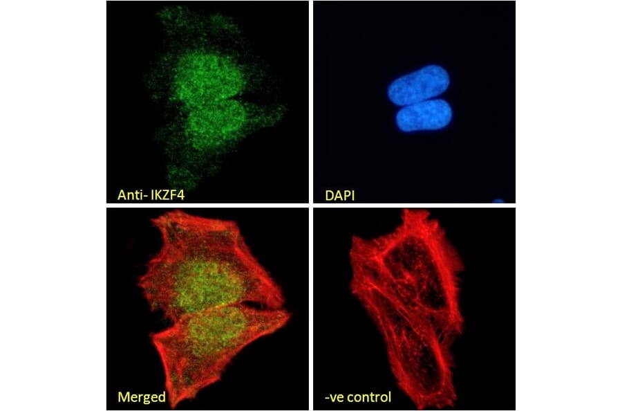
IKZF4 expression in HeLa cells analyzed by immunofluorescence. Cells were permeabilized with 0.15% Triton. Staining was performed with Anti-IKZF4 Antibody (A326255) at 10µg/ml for 1 hour and Alexa Fluor 488 secondary antibody at 2µg/ml. Nuclear staining shown and nuclei were stained with DAPI (blue) while actin filaments were stained with phalloidin (red). Negative control: Goat IgG Isotype Control at 10µg/ml followed by Alexa Fluor 488 secondary antibody at 2µg/ml.
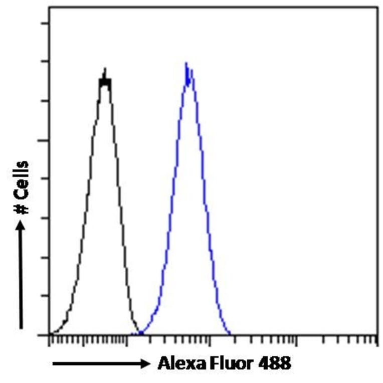
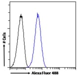
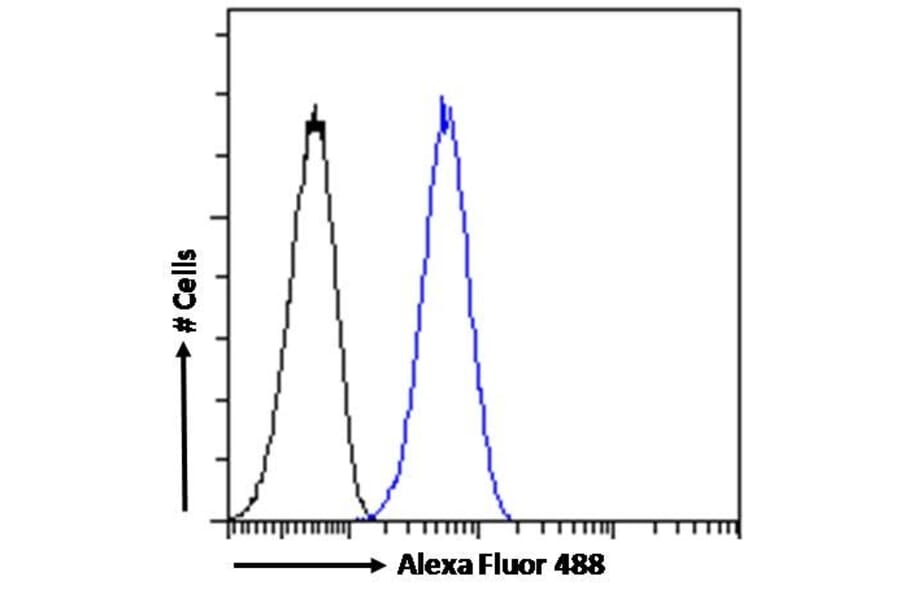
IKZF4 expression in HeLa cells (blue line) analyzed by flow cytometry. Cells were fixed in PFA and permeabilized with 0.5% Triton. Staining was performed with Anti-IKZF4 Antibody (A326255) at 10µg/ml for 1 hour and Alexa Fluor 488 secondary antibody at 1µg/ml. Negative Control: Goat IgG Isotype Control (black line) followed by Alexa Fluor 488 secondary antibody.