This antibody reacts with Linker for activation of T cells (LAT), a 36-38 kDa transmembrane adaptor protein expressed by T cells, pre-B cells, NK cells, mast cells and platelets.
Applications
WB, IP, Flow Cytometry (Intracellular)
Dilutions
IP: Tyrosine-phosphorylated form of LAT is not optimally recognized, Flow Cytometry: 1-4 µg/ml; Intracellular staining. Tyrosine-phosphorylated form of LAT is not optimally recognized, WB: 1-2 µg/ml.
Reactivity
Human
Cross Reactivity
This antibody does not cross react with Mouse.
Immunogen
Bacterially produced recombinant polypeptide corresponding to the entire cytoplasmic domain of human LAT.
Host
Mouse
Clonality
Monoclonal
Clone ID
LAT-01
Isotype
IgG1
Conjugate
Unconjugated
Purification
Protein A chromatography.
Concentration
1 mg/ml
Predicted MW
36-38 kDa
Product Form
Liquid
Formulation
Supplied in Phosphate Buffered Saline, pH 7.4, with 15 mM Sodium Azide.
Storage
Shipped at 4°C. Upon delivery aliquot and store at -20°C. Avoid freeze/thaw cycles.
Synonyms
36 kDa phosphotyrosine adapter protein, Linker for activation of T-cells family member 1, p36-38, pp36
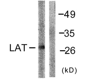
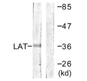
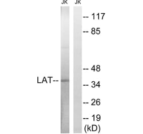
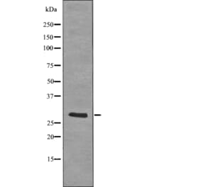
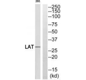
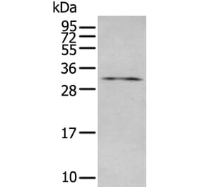
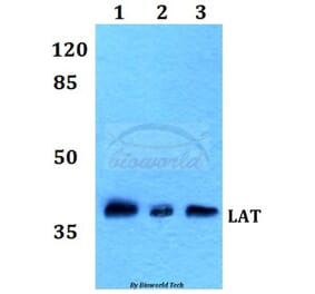
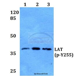
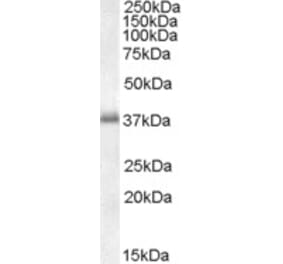
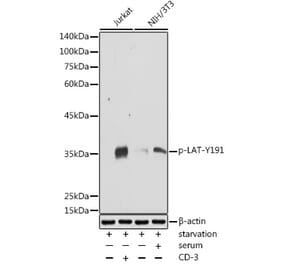
Western blotting analysis of human LAT using Anti-LAT Antibody [LAT-01] (A86328)in lysates of Jurkat cells (positive) and Raji cells (negative control) under non-reducing and reducing conditions. Nitrocellulose membrane was probed with 2 µg/ml of mouse anti-LAT monoclonal antibody followed by IRDye800-conjugated anti-mouse secondary antibody. A specific band was detected for LAT at approximately 38 kDa.
Separation of human CD3 positive LAT positive lymphocytes (red-filled) from neutrophil granulocytes (black-dashed) in flow cytometry analysis (intracellular staining) of peripheral whole blood stained with Anti-LAT Antibody [LAT-01] (A86328), (concentration in sample 1 µg/ml, GAM APC).


![WB - Anti-LAT Antibody [LAT-01] (A86328)](https://cdn.antibodies.com/image/catalog/86/A86328_1.jpg?profile=product_top)
![Flow Cytometry - Anti-LAT Antibody [LAT-01] (A86328)](https://cdn.antibodies.com/image/catalog/86/A86328_2.jpg?profile=product_top)
![Flow Cytometry - Anti-LAT Antibody [LAT-01] (A86328)](https://cdn.antibodies.com/image/catalog/86/A86328_3.jpg?profile=product_top)
![Flow Cytometry - Anti-LAT Antibody [LAT-01] (A86328)](https://cdn.antibodies.com/image/catalog/86/A86328_4.jpg?profile=product_top)
![WB - Anti-LAT Antibody [LAT-01] (A86328)](https://cdn.antibodies.com/image/catalog/86/A86328_1.jpg?profile=product_top_thumb)
![Flow Cytometry - Anti-LAT Antibody [LAT-01] (A86328)](https://cdn.antibodies.com/image/catalog/86/A86328_2.jpg?profile=product_top_thumb)
![Flow Cytometry - Anti-LAT Antibody [LAT-01] (A86328)](https://cdn.antibodies.com/image/catalog/86/A86328_3.jpg?profile=product_top_thumb)
![Flow Cytometry - Anti-LAT Antibody [LAT-01] (A86328)](https://cdn.antibodies.com/image/catalog/86/A86328_4.jpg?profile=product_top_thumb)
![WB - Anti-LAT Antibody [LAT-01] (A86328)](https://cdn.antibodies.com/image/catalog/86/A86328_1.jpg?profile=product_image)
![Flow Cytometry - Anti-LAT Antibody [LAT-01] (A86328)](https://cdn.antibodies.com/image/catalog/86/A86328_2.jpg?profile=product_image)
![Flow Cytometry - Anti-LAT Antibody [LAT-01] (A86328)](https://cdn.antibodies.com/image/catalog/86/A86328_3.jpg?profile=product_image)
![Flow Cytometry - Anti-LAT Antibody [LAT-01] (A86328)](https://cdn.antibodies.com/image/catalog/86/A86328_4.jpg?profile=product_image)