Immunocytochemistry/Immunofluorescence analysis of human neuroblastoma cells (SH-SY5Y), fixed in 4% PFA for 15 min, using Anti-Nav1.7 Antibody [N68/6] (A304801), at 1:100 for overnight at 4°C with slow rocking. The secondary antibody used was AlexaFluor 488 at 1:1,000 for 1 hour at room temperature. Counterstain: Phalloidin-iFluor 647 (red) F-Actin stain; Hoechst (blue) nuclear stain at 1:800, 1.6mM for 20 minutes at room temperature.(A) Hoechst (blue) nuclear stain. (B) Phalloidin-iFluor 647 (red) F-Actin stain. (C) Nav1.7 Antibody (D) Composite.
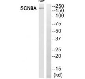
Western Blot - Anti-Nav1.7 Antibody [N68/6] (A304801)
Western blot analysis of Hamster CHO cells showing detection of Nav1.7 Sodium Channel protein using Anti-Nav1.7 Antibody [N68/6] (A304801) at 1:1,000 for 2 hours at room temperature. Load: 15 µg. Block: 1.5% BSA for 30 minutes at room temperature. The secondary antibody used was Sheep Anti-Mouse IgG: HRP for 1 hour at room temperature.
Immunohistochemistry analysis of mouse backskin, fixed in Bouin's fixative solution and paraffin-embedded. The Primary Antibody used was Anti-Nav1.7 Antibody [N68/6] (A304801) at 1:100 for 1 hour at room temperature. The secondary antibody used was FITC Goat Anti-Mouse (green) at 1:50 for 1 hour at room temperature.
Immunohistochemistry analysis of human hippocampus, fixed in Bouin's fixative solution and paraffin-embedded. The Primary Antibody used was Anti-Nav1.7 Antibody [N68/6] (A304801) at 1:1,000 for 1 hour at room temperature. The secondary antibody used was FITC Goat Anti-Mouse (green) at 1:50 for 1 hour at room temperature.
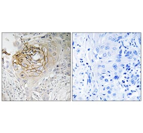
Immunohistochemistry analysis of mouse brain slice, fixed in frozen sections. The Primary Antibody used was Anti-Nav1.7 Antibody [N68/6] (A304801) at 1:1,000. The secondary antibody used was HRP/DAB Detection System: Biotinylated Goat Anti-Mouse, Streptavidin Peroxidase, DAB Chromogen (brown). Counterstain: Mayer Hematoxylin (purple/blue) nuclear stain.
Recombinant humanantibody to Nav17 for use as a research grade Duke anti-NAv17 biosimilar for ELISA, Flow Cytometry, Functional Studies and in vivo Research.
Recombinant humanantibody to Nav17 for use as a research grade Duke anti-NAv17 biosimilar for ELISA, Flow Cytometry, Functional Studies and in vivo Research.


![Immunocytochemistry/Immunofluorescence - Anti-Nav1.7 Antibody [N68/6] (A304801) - Antibodies.com](https://cdn.antibodies.com/image/catalog/304/A304801_1.png?profile=product_top)
![Western Blot - Anti-Nav1.7 Antibody [N68/6] (A304801) - Antibodies.com](https://cdn.antibodies.com/image/catalog/304/A304801_2.png?profile=product_top)
![Immunohistochemistry - Anti-Nav1.7 Antibody [N68/6] (A304801) - Antibodies.com](https://cdn.antibodies.com/image/catalog/304/A304801_3.png?profile=product_top)
![Immunohistochemistry - Anti-Nav1.7 Antibody [N68/6] (A304801) - Antibodies.com](https://cdn.antibodies.com/image/catalog/304/A304801_4.png?profile=product_top)
![Immunohistochemistry - Anti-Nav1.7 Antibody [N68/6] (A304801) - Antibodies.com](https://cdn.antibodies.com/image/catalog/304/A304801_5.png?profile=product_top)
![Immunocytochemistry/Immunofluorescence - Anti-Nav1.7 Antibody [N68/6] (A304801) - Antibodies.com](https://cdn.antibodies.com/image/catalog/304/A304801_1.png?profile=product_top_thumb)
![Western Blot - Anti-Nav1.7 Antibody [N68/6] (A304801) - Antibodies.com](https://cdn.antibodies.com/image/catalog/304/A304801_2.png?profile=product_top_thumb)
![Immunohistochemistry - Anti-Nav1.7 Antibody [N68/6] (A304801) - Antibodies.com](https://cdn.antibodies.com/image/catalog/304/A304801_3.png?profile=product_top_thumb)
![Immunohistochemistry - Anti-Nav1.7 Antibody [N68/6] (A304801) - Antibodies.com](https://cdn.antibodies.com/image/catalog/304/A304801_4.png?profile=product_top_thumb)
![Immunohistochemistry - Anti-Nav1.7 Antibody [N68/6] (A304801) - Antibodies.com](https://cdn.antibodies.com/image/catalog/304/A304801_5.png?profile=product_top_thumb)
![Immunocytochemistry/Immunofluorescence - Anti-Nav1.7 Antibody [N68/6] (A304801) - Antibodies.com](https://cdn.antibodies.com/image/catalog/304/A304801_1.png?profile=product_image)
![Western Blot - Anti-Nav1.7 Antibody [N68/6] (A304801) - Antibodies.com](https://cdn.antibodies.com/image/catalog/304/A304801_2.png?profile=product_image)
![Immunohistochemistry - Anti-Nav1.7 Antibody [N68/6] (A304801) - Antibodies.com](https://cdn.antibodies.com/image/catalog/304/A304801_3.png?profile=product_image)
![Immunohistochemistry - Anti-Nav1.7 Antibody [N68/6] (A304801) - Antibodies.com](https://cdn.antibodies.com/image/catalog/304/A304801_4.png?profile=product_image)
![Immunohistochemistry - Anti-Nav1.7 Antibody [N68/6] (A304801) - Antibodies.com](https://cdn.antibodies.com/image/catalog/304/A304801_5.png?profile=product_image)
![SDS-PAGE - Anti-Nav1.7 Antibody [Research Grade Biosimilar] - Low endotoxin, Azide free (A324160) - Antibodies.com](https://cdn.antibodies.com/image/catalog/324/A324160_1.jpg?profile=product_alternative)
![SDS-PAGE - Anti-Nav1.7 Antibody [Duke anti-NAv1.7] Biosimilar - BSA and Azide free (A340794) - Antibodies.com](https://cdn.antibodies.com/image/catalog/340/A340794_1.jpg?profile=product_alternative)