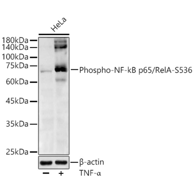
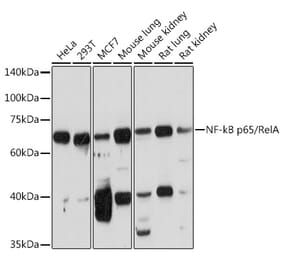
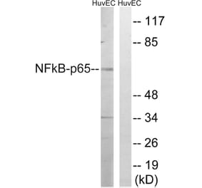
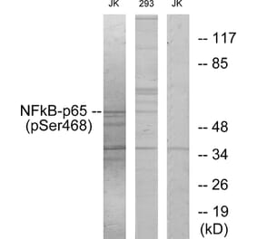
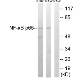
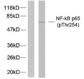
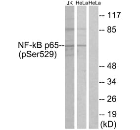
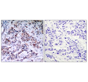
Western Blot - Anti-NF-kB p65 (phospho Ser536) Antibody (A91007)
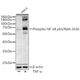
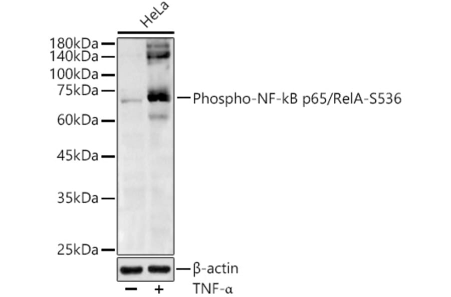
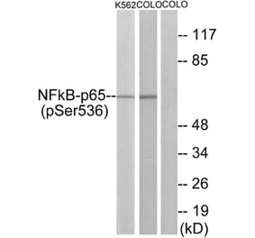
Western blot analysis of extracts of various cell lines, using Anti-NF-kB p65 (phospho Ser536) Antibody (A91007) at 1:1,000 dilution. The secondary antibody was Goat Anti-Rabbit IgG H&L Antibody (HRP) at 1:10,000 dilution. Lysates/proteins were present at 25µg per lane. The blocking buffer used was 3% non-fat dry milk in TBST. Detection was with a ECL Basic Kit. Exposure time: 60s.
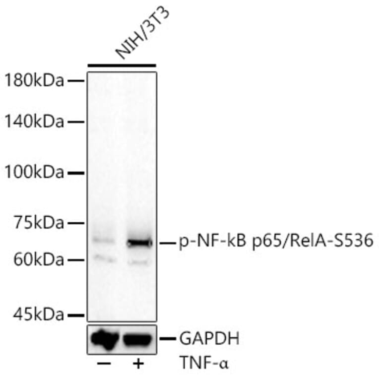
Western Blot - Anti-NF-kB p65 (phospho Ser536) Antibody (A91007)
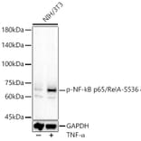
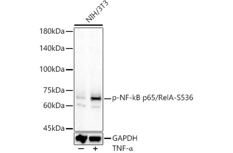
Western blot analysis of NIH/3T3, using Anti-NF-kB p65 (phospho Ser536) Antibody (A91007) at 1:600 dilution. NIH/3T3 cells were treated by TNF-alpha (20 ng/ml) at 37°C for 30 minutes. The secondary antibody was Goat Anti-Rabbit IgG H&L Antibody (HRP) at 1:10,000 dilution. Lysates/proteins were present at 25µg per lane. The blocking buffer used was 3% non-fat dry milk in TBST. Detection was with a ECL Basic Kit. Exposure time: 60s.
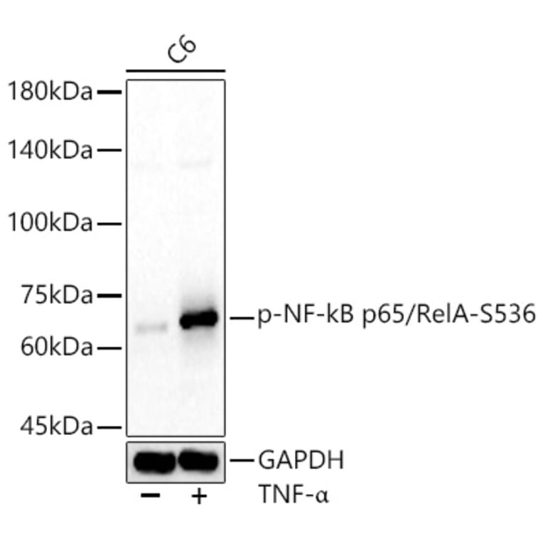
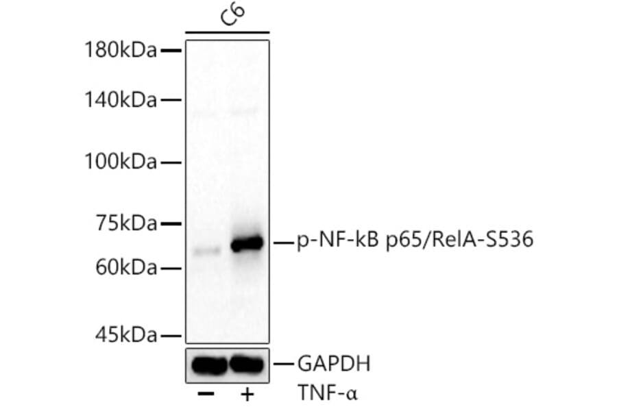
Western Blot - Anti-NF-kB p65 (phospho Ser536) Antibody (A91007)
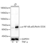
Western blot analysis of C6, using Anti-NF-kB p65 (phospho Ser536) Antibody (A91007) at 1:600 dilution. C6 cells were treated by TNF-alpha (20 ng/ml) at 37°C for 30 minutes. The secondary antibody was Goat Anti-Rabbit IgG H&L Antibody (HRP) at 1:10,000 dilution. Lysates/proteins were present at 25µg per lane. The blocking buffer used was 3% non-fat dry milk in TBST. Detection was with a ECL Basic Kit. Exposure time: 60s.
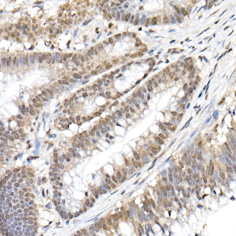
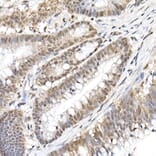
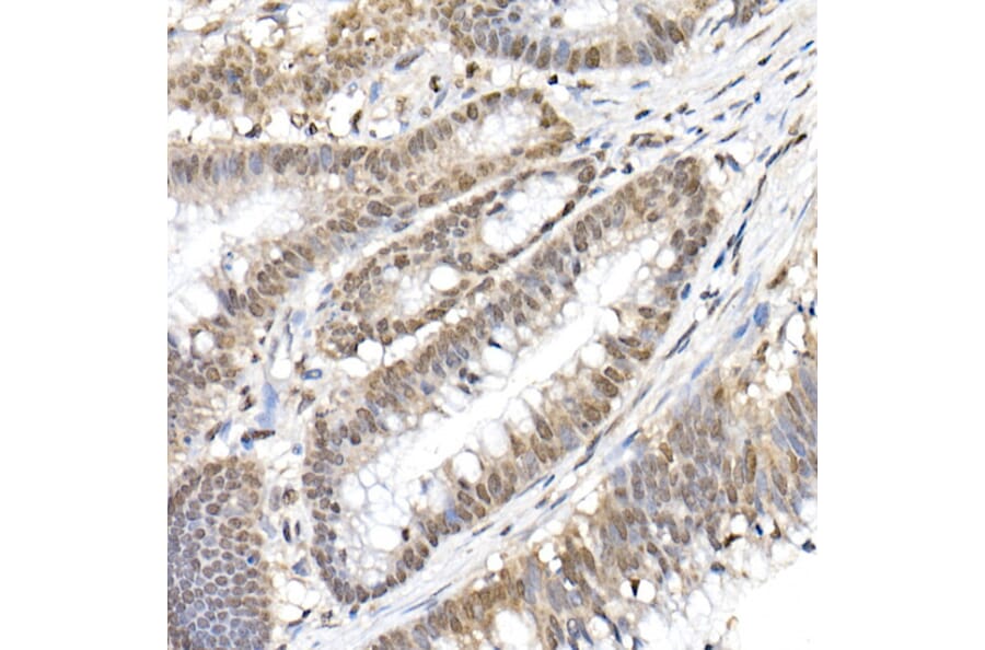
Immunohistochemistry analysis of paraffin-embedded human colon carcinoma tissue using Anti-NF-kB p65 (phospho Ser536) Antibody (A91007) at a dilution of 1:50 (40x lens). Perform high pressure antigen retrieval with 10 mM citrate buffer pH 6.0 before commencing with IHC staining protocol.
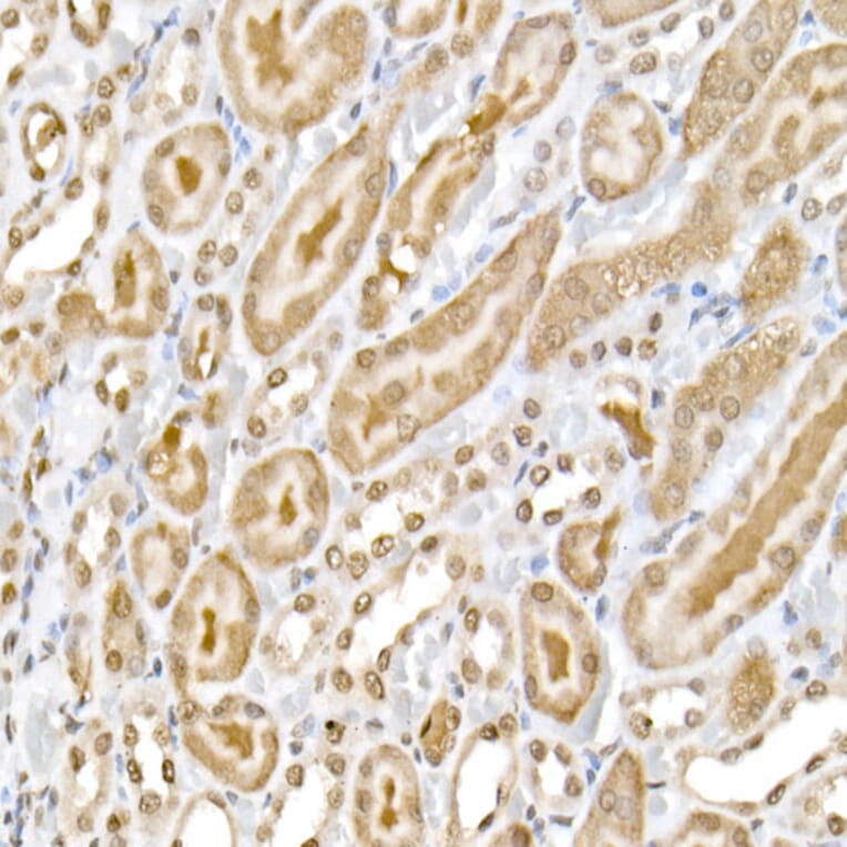
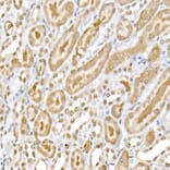
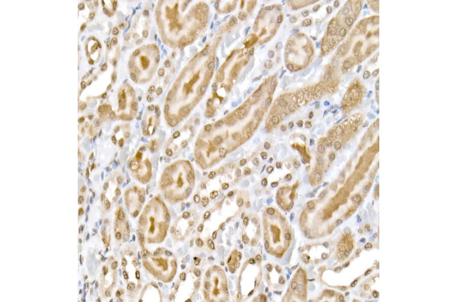
Immunohistochemistry analysis of paraffin-embedded rat kidney using Anti-NF-kB p65 (phospho Ser536) Antibody (A91007) at a dilution of 1:50 (40x lens). Perform high pressure antigen retrieval with 10 mM citrate buffer pH 6.0 before commencing with IHC staining protocol.