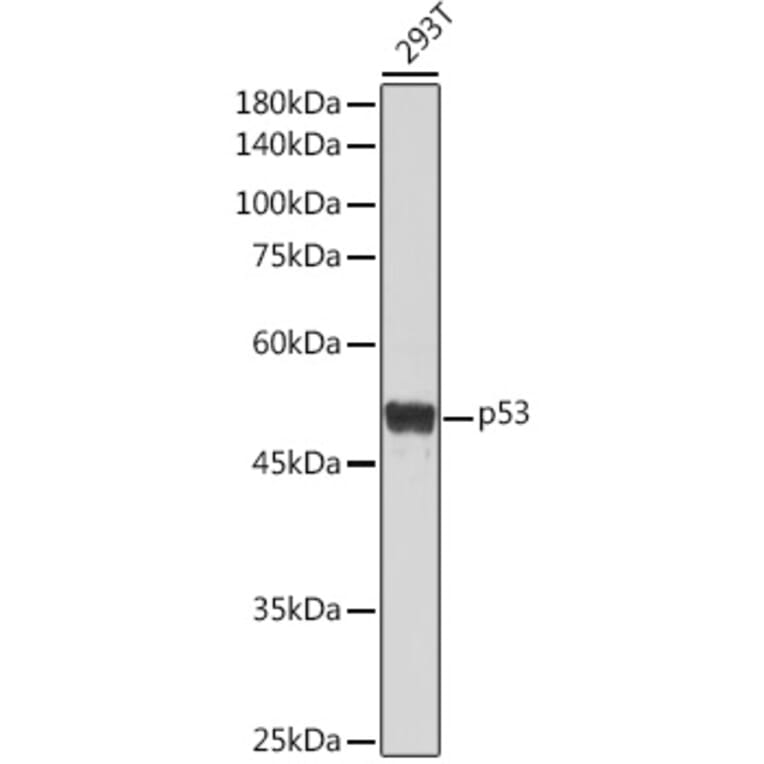
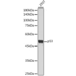
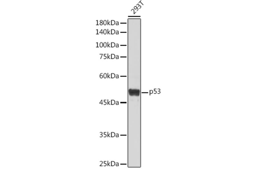
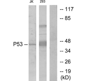
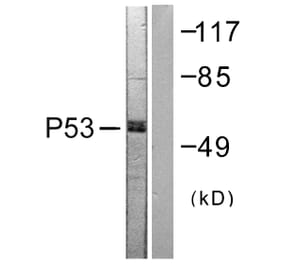
Western blot analysis of extracts of 293T cells, using Anti-p53 Antibody (A309435) at 1:1,000 dilution. The secondary antibody was Goat Anti-Rabbit IgG H&L Antibody (HRP) at 1:10,000 dilution. Lysates/proteins were present at 25µg per lane. The blocking buffer used was 3% non-fat dry milk in TBST. Detection was with a ECL Basic Kit. Exposure time: 30s.
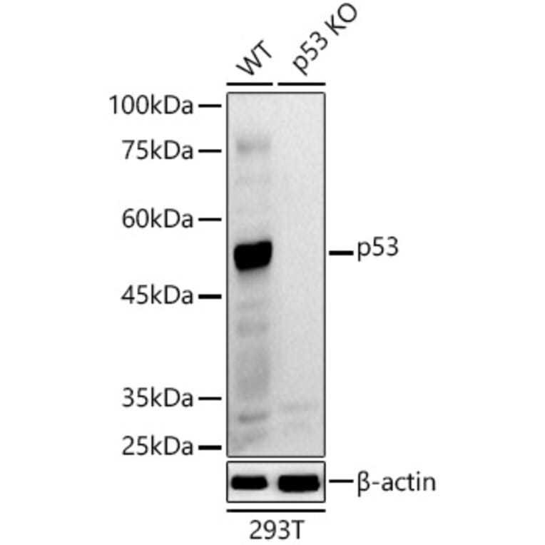
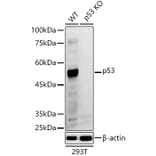
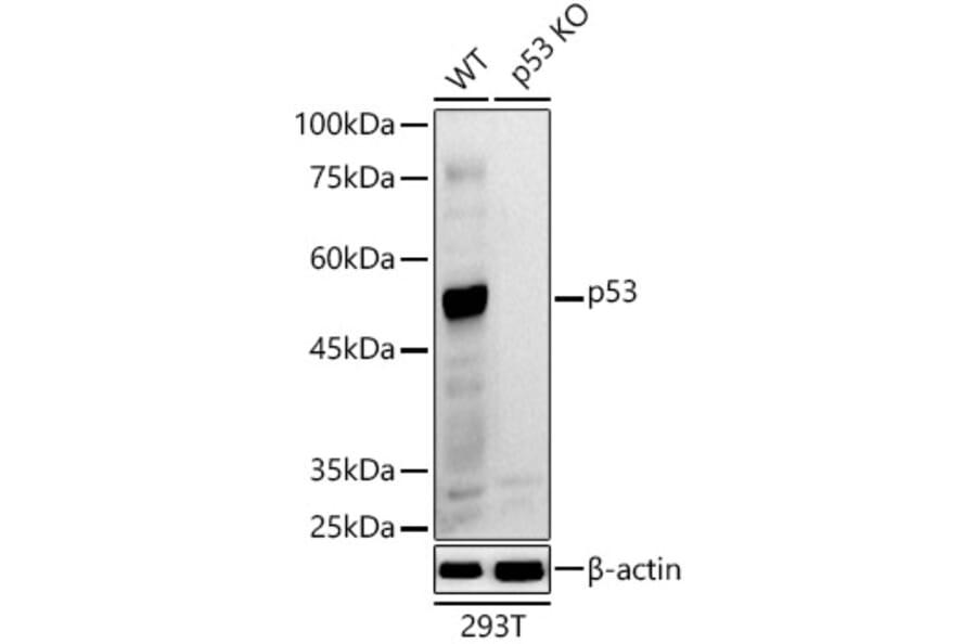
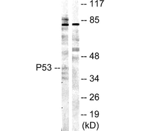
Western blot analysis of extracts from wild type (WT) and p53 knockout (KO) 293T cells, using Anti-p53 Antibody (A309435) at 1:1,000 dilution. The secondary antibody was Goat Anti-Rabbit IgG H&L Antibody (HRP) at 1:10,000 dilution. Lysates/proteins were present at 25µg per lane. The blocking buffer used was 3% non-fat dry milk in TBST. Detection was with a ECL Basic Kit. Exposure time: 90s.
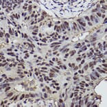
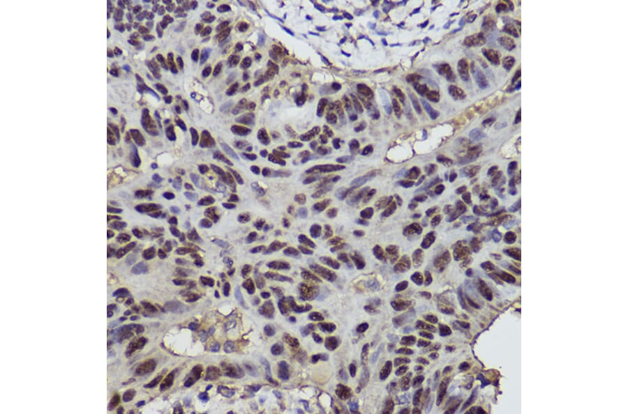
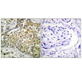
Immunohistochemistry analysis of paraffin-embedded human colon carcinoma tissue using Anti-p53 Antibody (A309435) at a dilution of 1:300 (40x lens). Perform high pressure antigen retrieval with 10 mM citrate buffer pH 6.0 before commencing with IHC staining protocol.
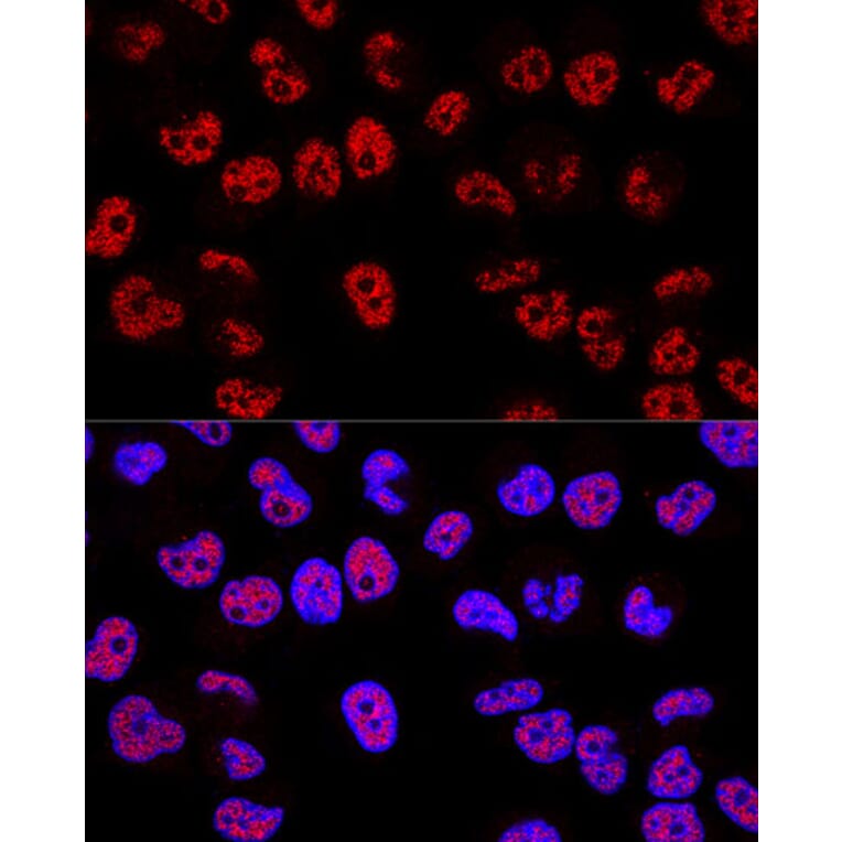
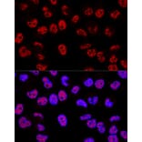
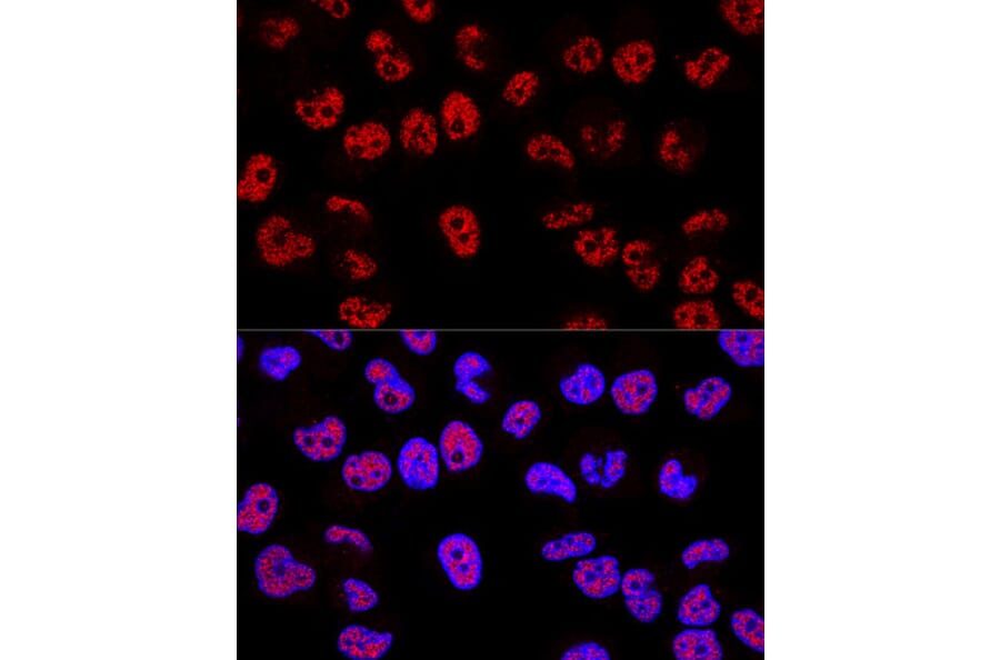
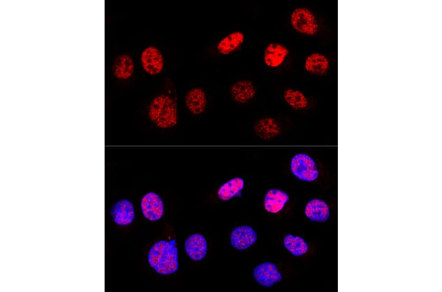
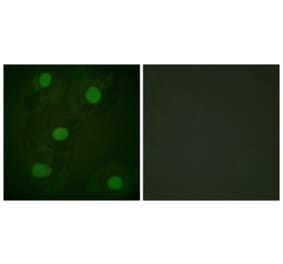
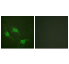
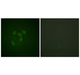
Confocal immunofluorescence analysis of Hela cells using Anti-p53 Antibody (A309435) at a dilution of 1:200. DAPI was used to stain the cell nuclei (blue).
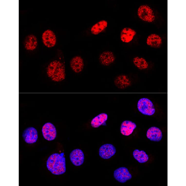
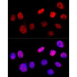
Confocal immunofluorescence analysis of U-2 OS cells using Anti-p53 Antibody (A309435) at a dilution of 1:200. DAPI was used to stain the cell nuclei (blue).
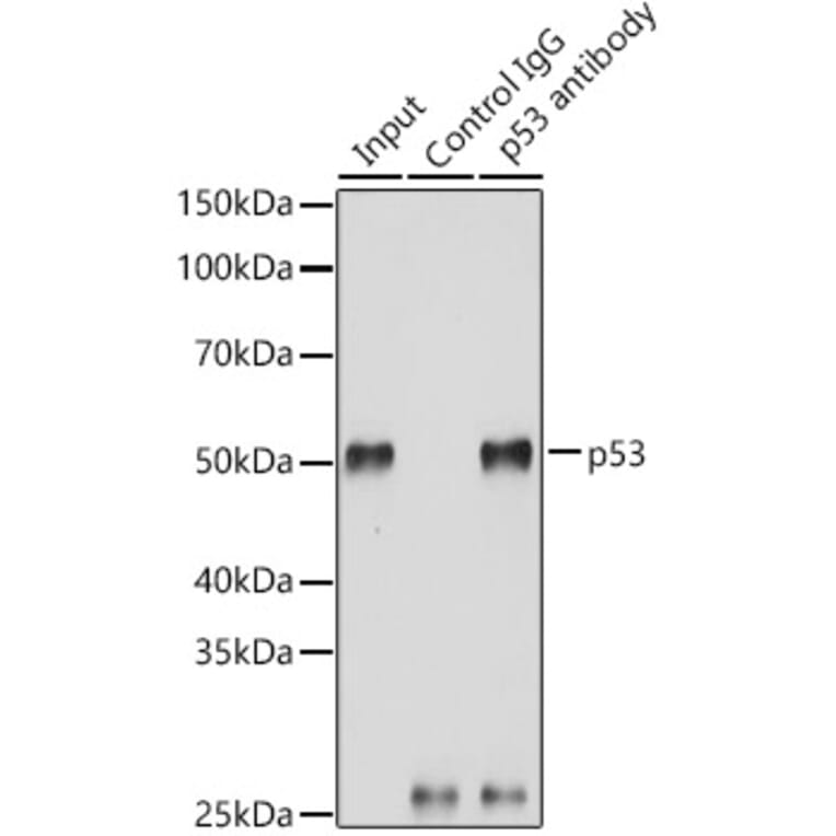
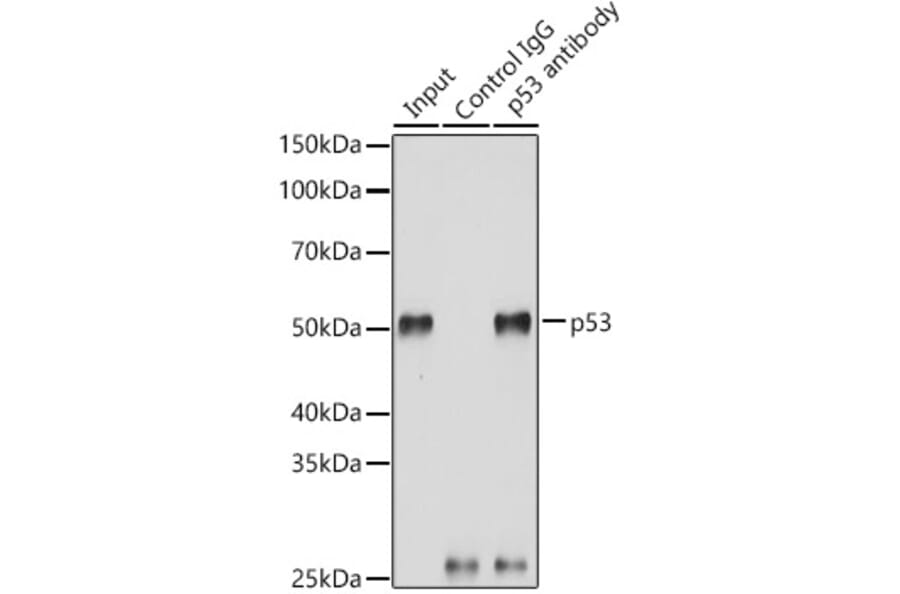
Immunoprecipitation analysis of 200µg extracts of 293T cells using 3µg of Anti-p53 Antibody (A309435). This Western blot was performed on the immunoprecipitate using Anti-p53 Antibody (A309435) at a dilution of 1:3000.
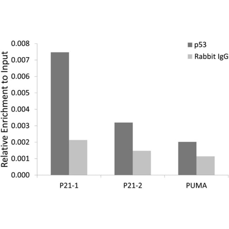
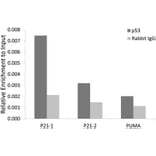
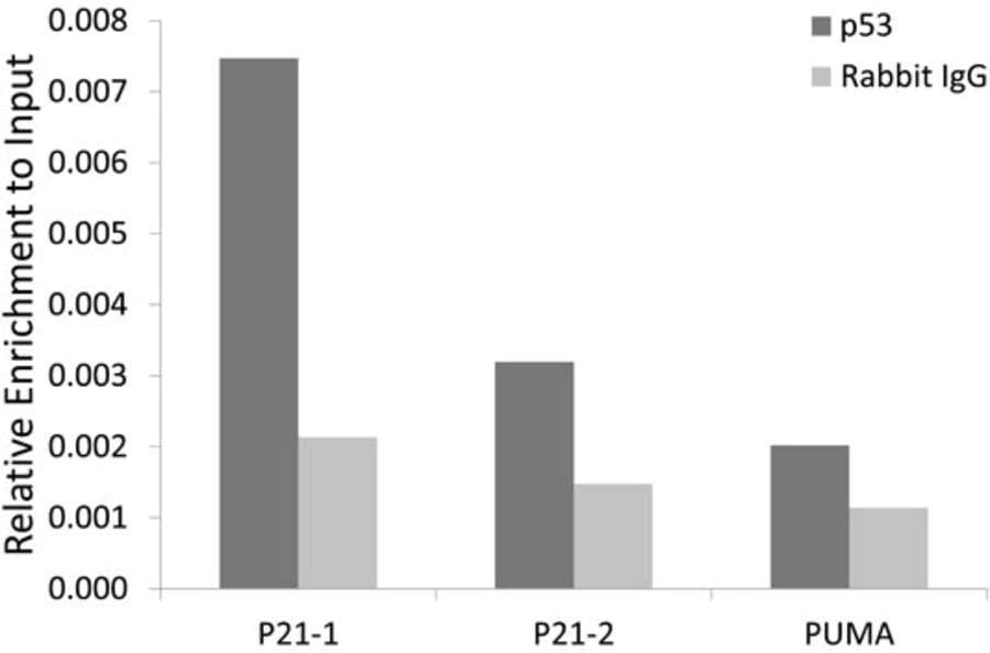
Chromatin immunoprecipitation (ChIP) analysis of extracts of 293T cells, using Anti-p53 Antibody (A309435) and Rabbit IgG. The amount of immunoprecipitated DNA was checked by quantitative PCR. Histogram was constructed by the ratios of the immunoprecipitated DNA to the input.