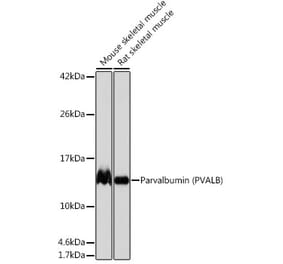
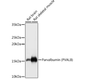
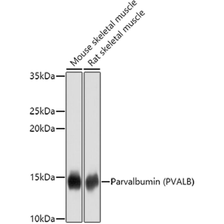
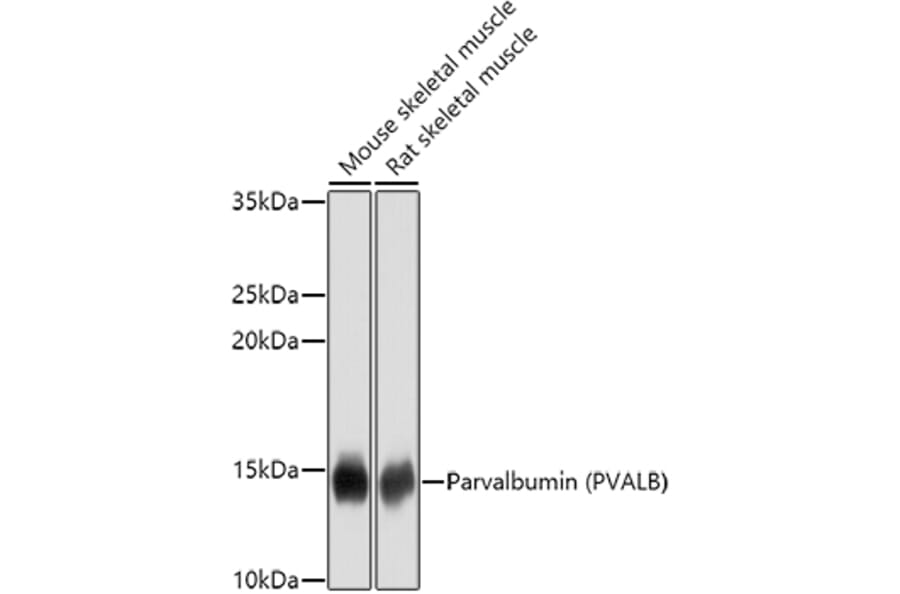
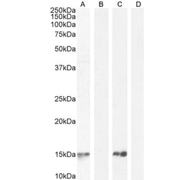
Parvalbumin expression in various lysates analyzed by western blot. Primary antibody incubation was performed for 1 hour on 25ug protein per lane with Anti-Parvalbumin Antibody (A14195) at a dilution of 1:1000 and detected with chemiluminescence.
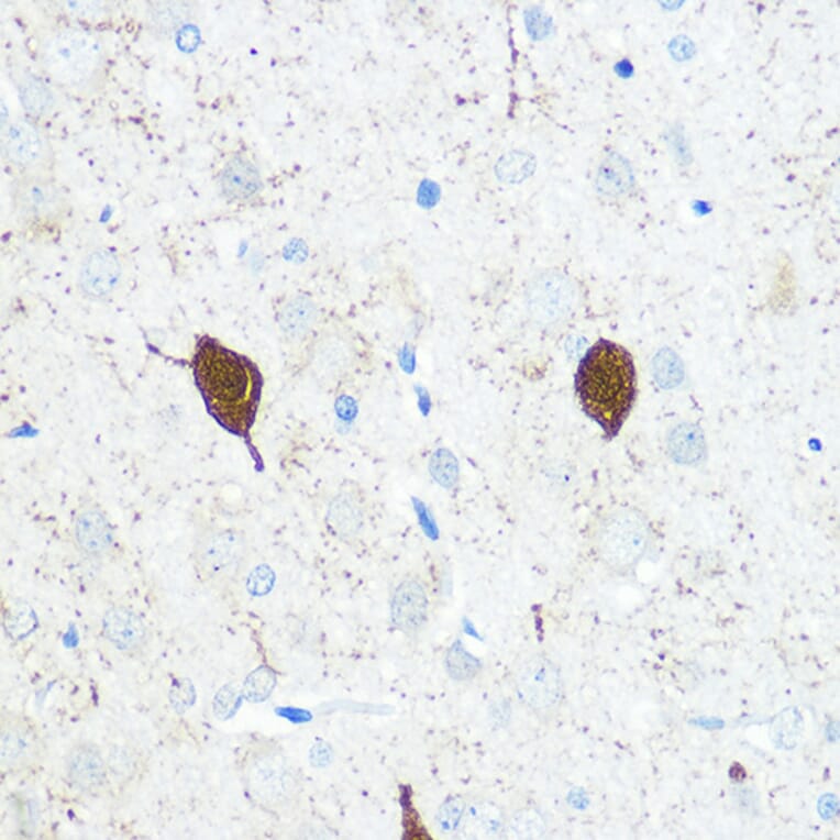
Parvalbumin expression in rat brain tissue analyzed by immunohistochemistry. Tissue was paraffin-embedded, and antigen retrieval was achieved by microwaving in 10 mM PBS buffer, pH 7.2. Staining was performed with Anti-Parvalbumin Antibody (A14195) at a dilution of 1:100.
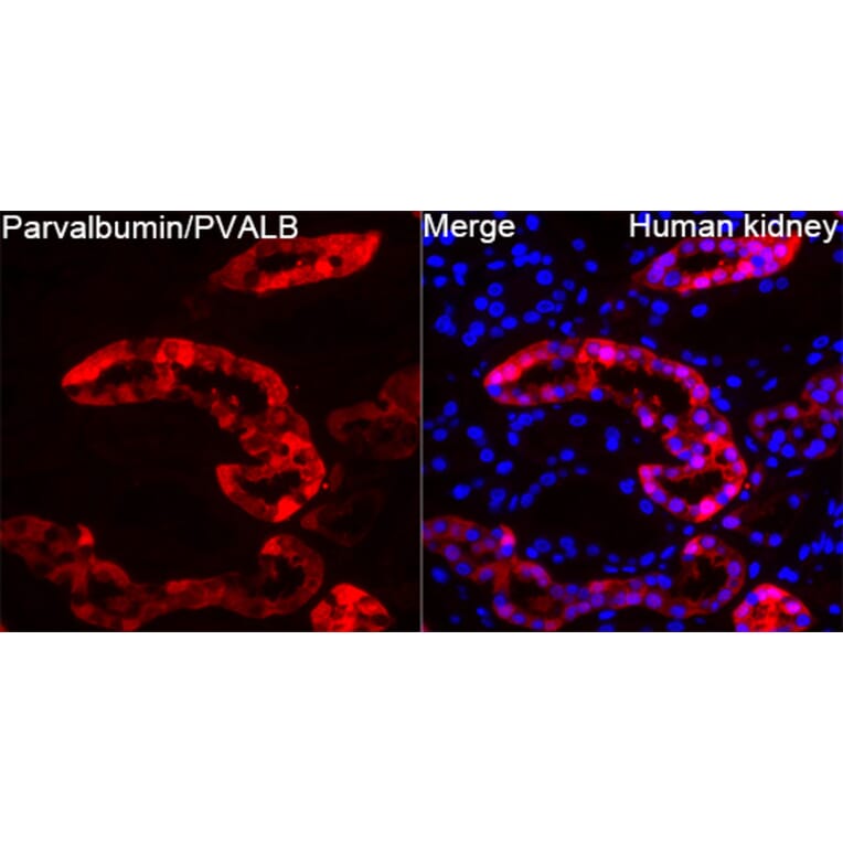
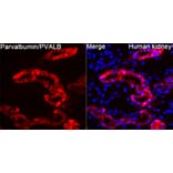
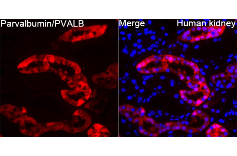
Parvalbumin expression in Human kidney tissue analyzed by immunofluorescence. Staining was performed with Anti-Parvalbumin Antibody (A14195) at a dilution of 1:100 followed by Cy3-conjugated Goat anti-Rabbit IgG (H+L)secondary antibody at a dilution of 1:500. Nuclei were stained with DAPI (blue).
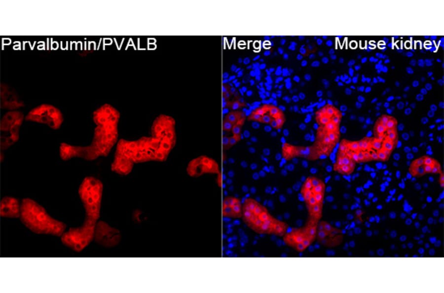
Parvalbumin expression in Mouse kidney tissue analyzed by immunofluorescence. Staining was performed with Anti-Parvalbumin Antibody (A14195) at a dilution of 1:100 followed by Cy3-conjugated Goat anti-Rabbit IgG (H+L)secondary antibody at a dilution of 1:500. Nuclei were stained with DAPI (blue).
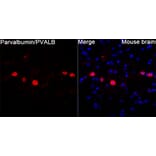
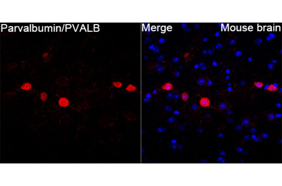
Parvalbumin expression in Mouse brain tissue analyzed by immunofluorescence. Staining was performed with Anti-Parvalbumin Antibody (A14195) at a dilution of 1:100 followed by Cy3-conjugated Goat anti-Rabbit IgG (H+L)secondary antibody at a dilution of 1:500. Nuclei were stained with DAPI (blue).
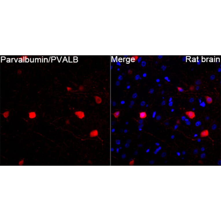
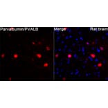
Parvalbumin expression in Rat brain tissue analyzed by immunofluorescence. Staining was performed with Anti-Parvalbumin Antibody (A14195) at a dilution of 1:100 followed by Cy3-conjugated Goat anti-Rabbit IgG (H+L)secondary antibody at a dilution of 1:500. Nuclei were stained with DAPI (blue).


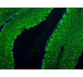
![Immunofluorescence - Anti-Parvalbumin Antibody [3C9] (A85317) - Antibodies.com](https://cdn.antibodies.com/image/catalog/85/A85317_1.jpg?profile=product_alternative)
![Immunohistochemistry - Anti-Parvalbumin Antibody [C12] (A305173) - Antibodies.com](https://cdn.antibodies.com/image/catalog/305/A305173_1.png?profile=product_alternative)