Supplied in Phosphate Buffered Saline, pH 7.3, with 50% Glycerol, 0.05% BSA, and 0.02% Sodium Azide.
Storage
Shipped at 4°C. Upon delivery aliquot and store at -20°C. Avoid freeze/thaw cycles.
Synonyms
Endoplasmic reticulum protein 5, ER protein 5, ERP5, P5, Protein disulfide isomerase P5, Protein disulfide-isomerase A6, Thioredoxin domain-containing protein 7, TXNDC7
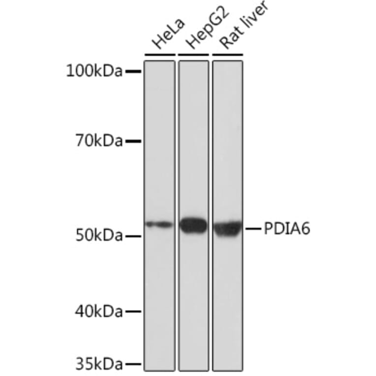
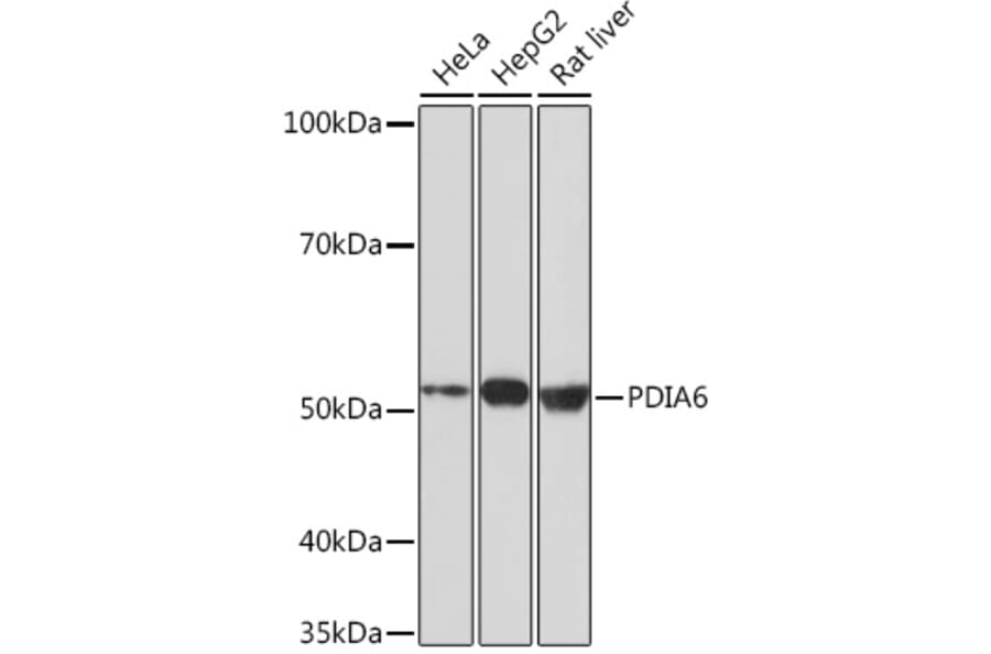
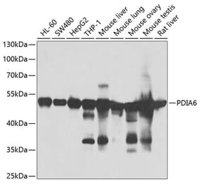
Western Blot - Anti-PDIA6 Antibody [ARC0944] (A306932)
Western blot analysis of extracts of various cell lines, using Anti-PDIA6 Antibody (A306932) at 1:1,000 dilution. The secondary antibody was Goat Anti-Rabbit IgG H&L Antibody (HRP) at 1:10,000 dilution. Lysates/proteins were present at 25µg per lane. The blocking buffer used was 3% non-fat dry milk in TBST. Detection was with a ECL Basic Kit. Exposure time: 3min.
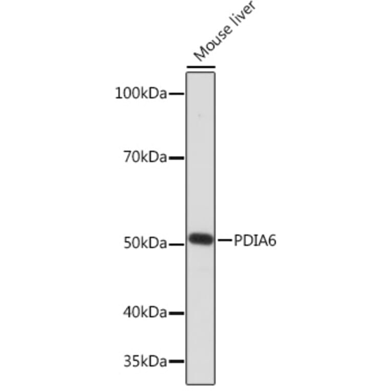
Western Blot - Anti-PDIA6 Antibody [ARC0944] (A306932)
Western blot analysis of extracts of Mouse liver, using Anti-PDIA6 Antibody (A306932) at 1:1,000 dilution. The secondary antibody was Goat Anti-Rabbit IgG H&L Antibody (HRP) at 1:10,000 dilution. Lysates/proteins were present at 25µg per lane. The blocking buffer used was 3% non-fat dry milk in TBST. Detection was with a ECL Enhanced Kit. Exposure time: 3min.
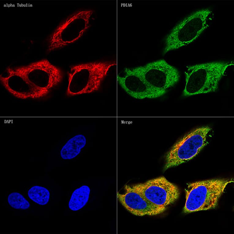
Confocal imaging of HeLa cells using Anti-PDIA6 Antibody (A306932), at a dilution of 1:100, (green). The cells were counterstained with Anti-alpha Tubulin Antibody, at a dilution of 1:100, (red). DAPI was used for nuclear staining (Blue). Objective: 60x.
Publishing research using Anti-PDIA6 Antibody [ARC0944] (A306932)? Please let us know so that we can list the citation on this page.
Alternative products to Anti-PDIA6 Antibody [ARC0944] (A306932)