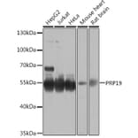
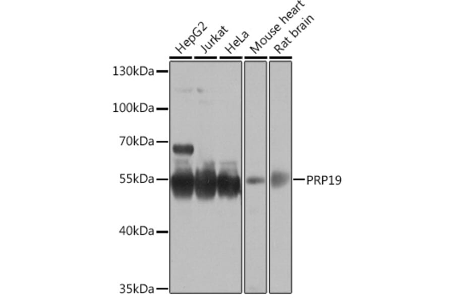
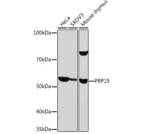
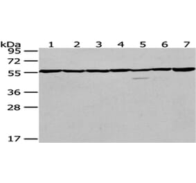
Western blot analysis of extracts of various cell lines, using Anti-PRP19 Antibody (A90482) at 1:1,000 dilution. The secondary antibody was Goat Anti-Rabbit IgG H&L Antibody (HRP) at 1:10,000 dilution. Lysates/proteins were present at 25µg per lane. The blocking buffer used was 3% non-fat dry milk in TBST. Detection was with a ECL Basic Kit. Exposure time: 30s.
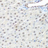
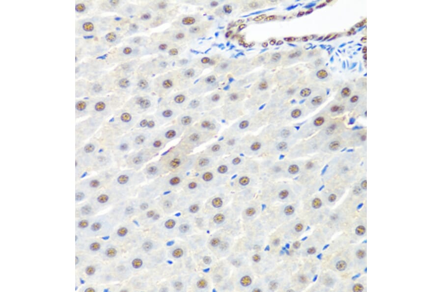
Immunohistochemistry analysis of paraffin-embedded rat liver using Anti-PRP19 Antibody (A90482) at a dilution of 1:100 (40x lens). Perform microwave antigen retrieval with 10 mM PBS buffer pH 7.2 before commencing with IHC staining protocol.
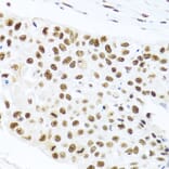
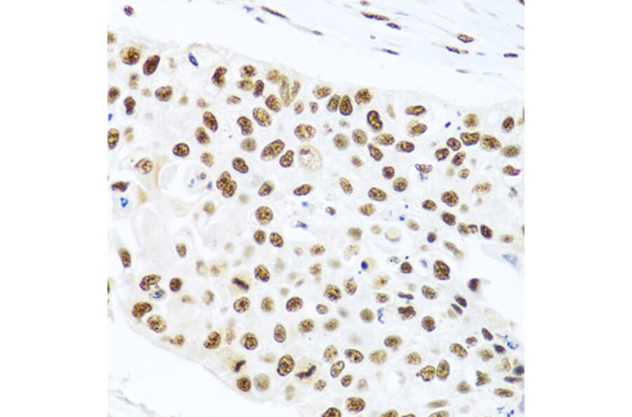
Immunohistochemistry analysis of paraffin-embedded human lung cancer using Anti-PRP19 Antibody (A90482) at a dilution of 1:100 (40x lens). Perform microwave antigen retrieval with 10 mM PBS buffer pH 7.2 before commencing with IHC staining protocol.
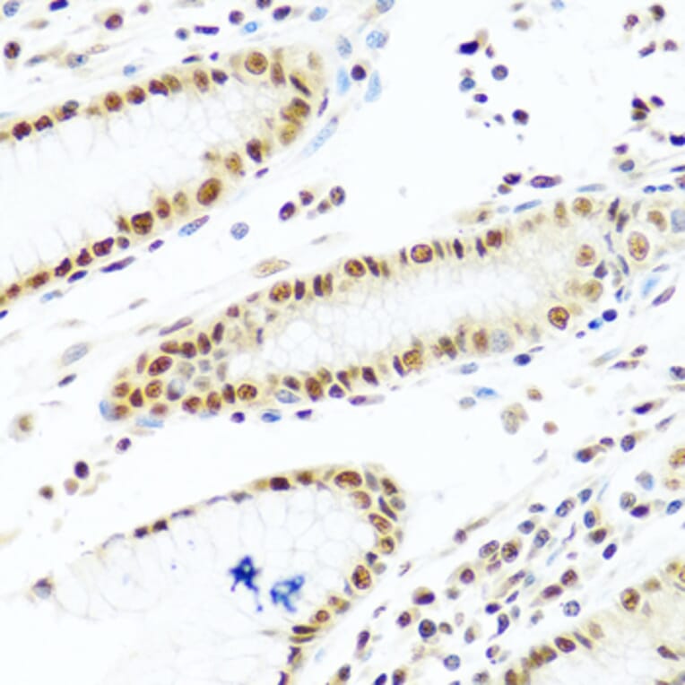
Immunohistochemistry analysis of paraffin-embedded human stomach using Anti-PRP19 Antibody (A90482) at a dilution of 1:100 (40x lens). Perform microwave antigen retrieval with 10 mM PBS buffer pH 7.2 before commencing with IHC staining protocol.
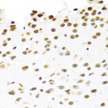
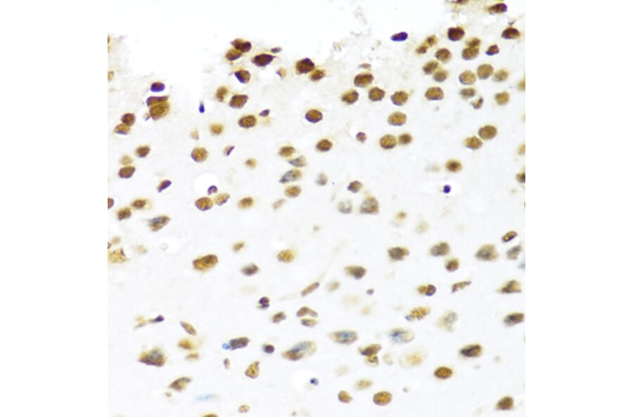
Immunohistochemistry analysis of paraffin-embedded mouse brain using Anti-PRP19 Antibody (A90482) at a dilution of 1:100 (40x lens). Perform microwave antigen retrieval with 10 mM PBS buffer pH 7.2 before commencing with IHC staining protocol.
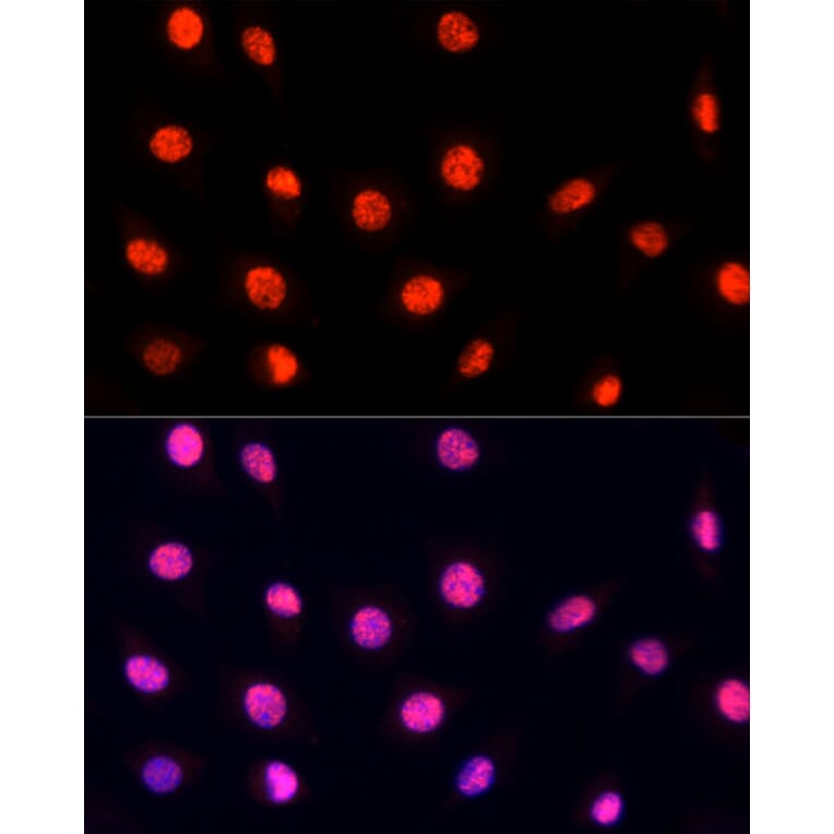
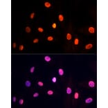
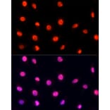
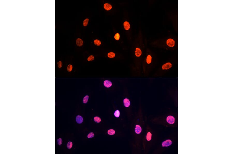
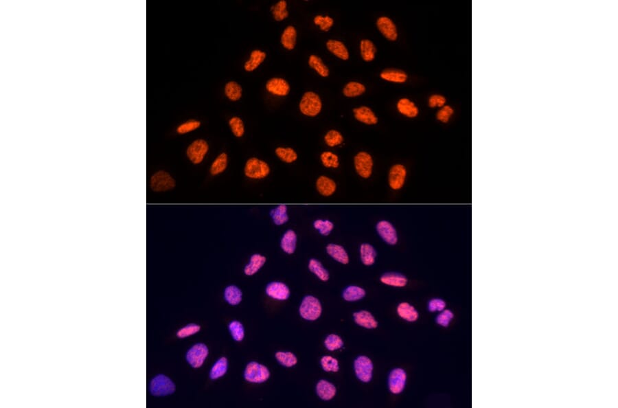
Immunofluorescence analysis of H9C2 cells using Anti-PRP19 Antibody (A90482) at a dilution of 1:100 (40x lens). DAPI was used to stain the cell nuclei (blue).
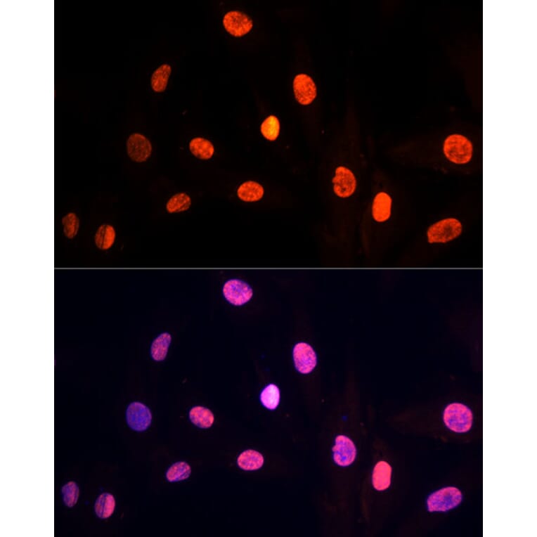
Immunofluorescence analysis of L929 cells using Anti-PRP19 Antibody (A90482) at a dilution of 1:100 (40x lens). DAPI was used to stain the cell nuclei (blue).
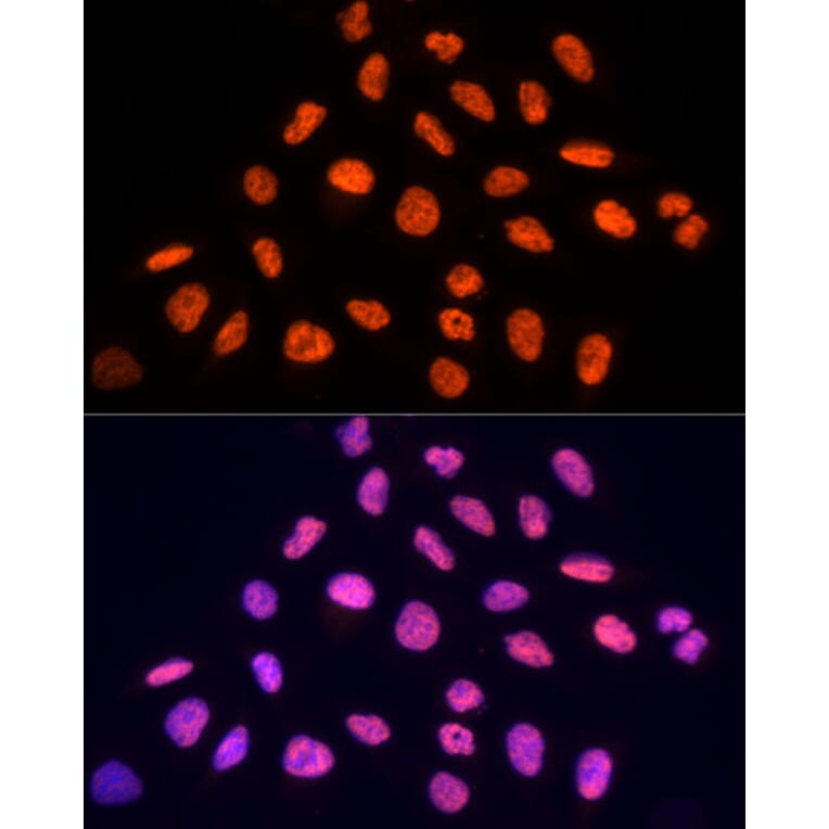
Immunofluorescence analysis of U2OS cells using Anti-PRP19 Antibody (A90482) at a dilution of 1:100 (40x lens). DAPI was used to stain the cell nuclei (blue).
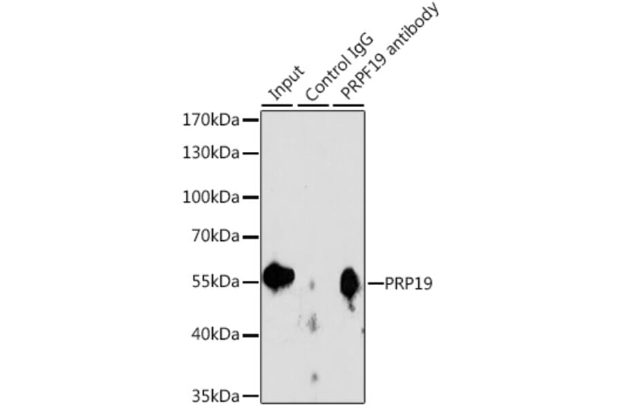
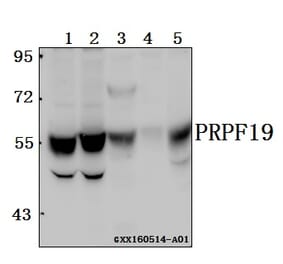
Immunoprecipitation analysis of 200µg extracts of Jurkat cells using 3µg of Anti-PRP19 Antibody (A90482). This Western blot was performed on the immunoprecipitate using Anti-PRP19 Antibody (A90482) at a dilution of 1:1000.