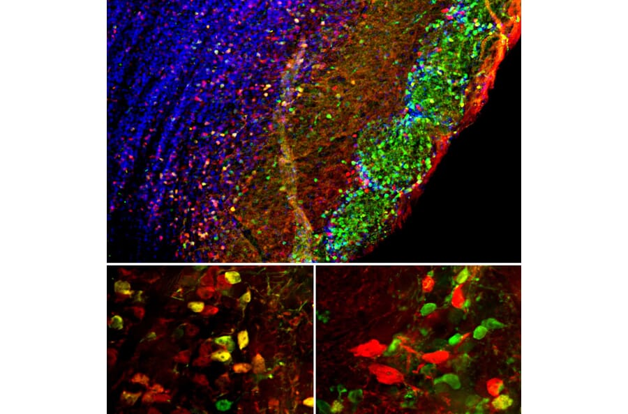
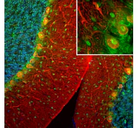
Immunofluorescent analysis of mouse brain olfactory bulb section stained with Anti-Secretagogin Antibody (A104317), at a dilution of 1:1,000, in red, and co-stained with Anti-Calretinin Antibody [3G9] (A85367), at a dilution of 1:500, in green. The blue is Hoechst staining of nuclear DNA. Following transcardial perfusion with 4% paraformaldehyde, brain was post fixed for 24 hours, cut to 45µM, and free-floating sections were stained with the above antibodies. Secretagogin is highly expressed in the mitral and granular cell layers, while Calretinin is in the glomerular layer. Cells that express secretagogin or calretinin only are red or green respectively. A number of cells containing both proteins appears orange-yellow.
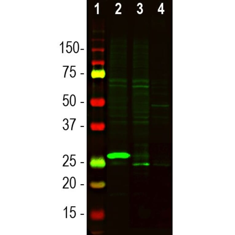
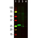
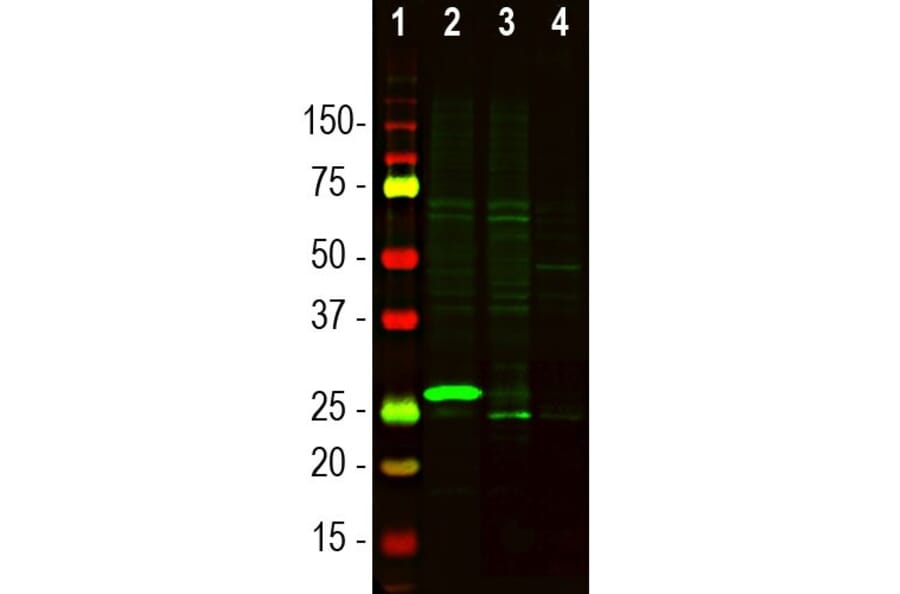
Western Blot - Anti-Secretagogin Antibody (A104317)
Western blot analysis of tissue lysates using Anti-Secretagogin Antibody (A104317), at a dilution of 1:1,000, in green. The lanes contain: [Lane 1] protein standard, [Lane 2] mouse olfactory bulb, [Lane 3] rat cerebellum, and [Lane 4] cow cerebellum. The band at ~27kDa corresponds to the Secretagogin protein. Secretagogin protein is highly expressed in the mouse olfactory bulb, but significantly less in the cerebellum.
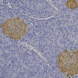
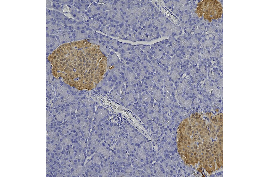
Immunohistochemistry analysis of a formalin fixed paraffin embedded mouse pancreas section with Anti-Secretagogin Antibody (A104317) at a dilution of 1:2,000 detected in DAB (brown) following the ABC method. Counterstained with Hematoxylin (blue). Anti-Secretagogin Antibody (A104317) robustly stains the alpha and beta cells of pancreatic islets.
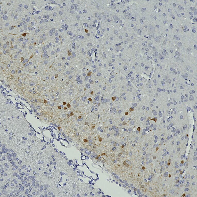
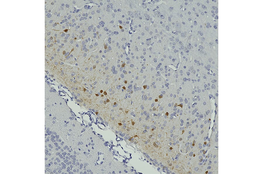
Immunohistochemistry analysis of a formalin fixed paraffin embedded rat brain section (adjacent to the lateral ventricle) with Anti-Secretagogin Antibody (A104317) at a dilution of 1:2,000 detected in DAB (brown) following the ABC method. Counterstained with Hematoxylin (blue).
Publishing research using Anti-Secretagogin Antibody (A104317)? Please let us know so that we can list the citation on this page.
Alternative products to Anti-Secretagogin Antibody (A104317)