Supplied in Phosphate Buffered Saline, pH 7.3, with 50% Glycerol and 0.02% Sodium Azide.
Storage
Shipped at 4°C. Upon delivery aliquot and store at -20°C. Avoid freeze/thaw cycles.
Synonyms
SH2 domain protein C1, SHC-transforming protein 1, SHC-transforming protein 3, SHC-transforming protein A, SHC1, SHCA, Src homology 2 domain-containing-transforming protein C1
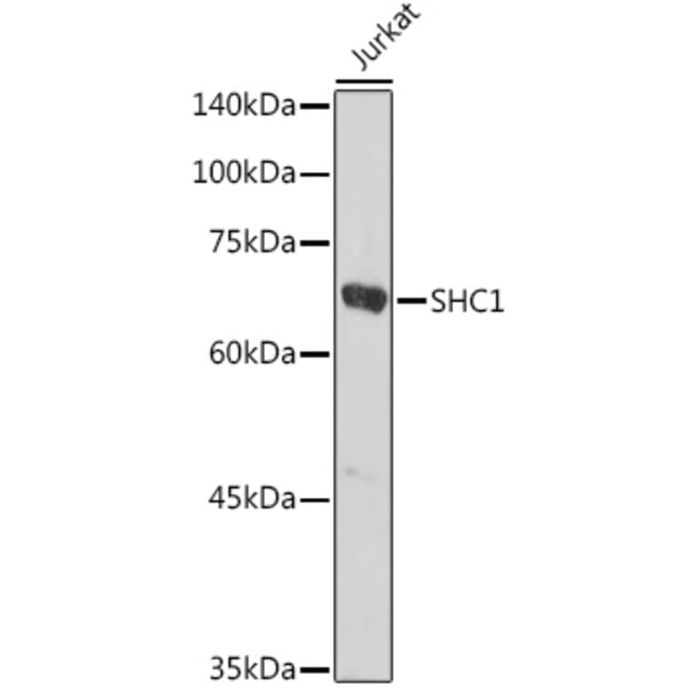
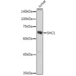
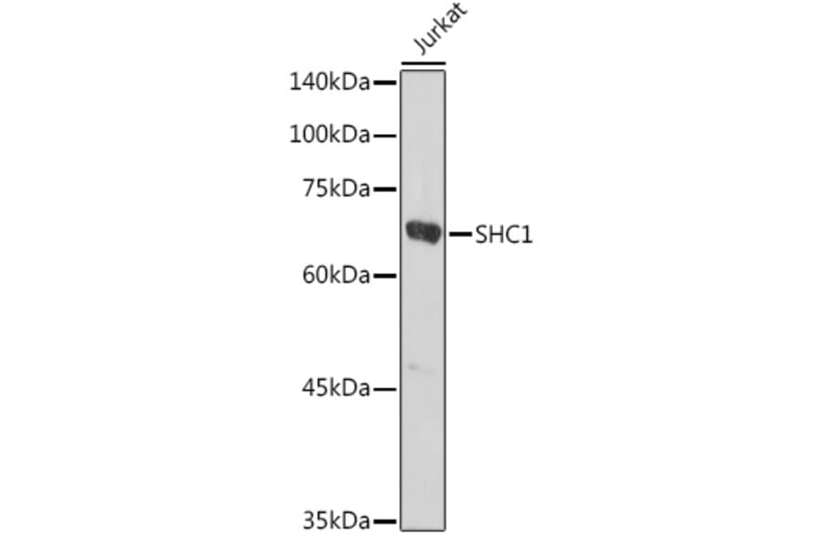
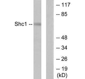
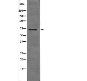
Western blot analysis of extracts of Jurkat cells, using Anti-SHC Antibody (A16912) at 1:1,000 dilution. The secondary antibody was Goat Anti-Rabbit IgG H&L Antibody (HRP) at 1:10,000 dilution. Lysates/proteins were present at 25µg per lane. The blocking buffer used was 3% non-fat dry milk in TBST. Detection was with a ECL Basic Kit. Exposure time: 30s.
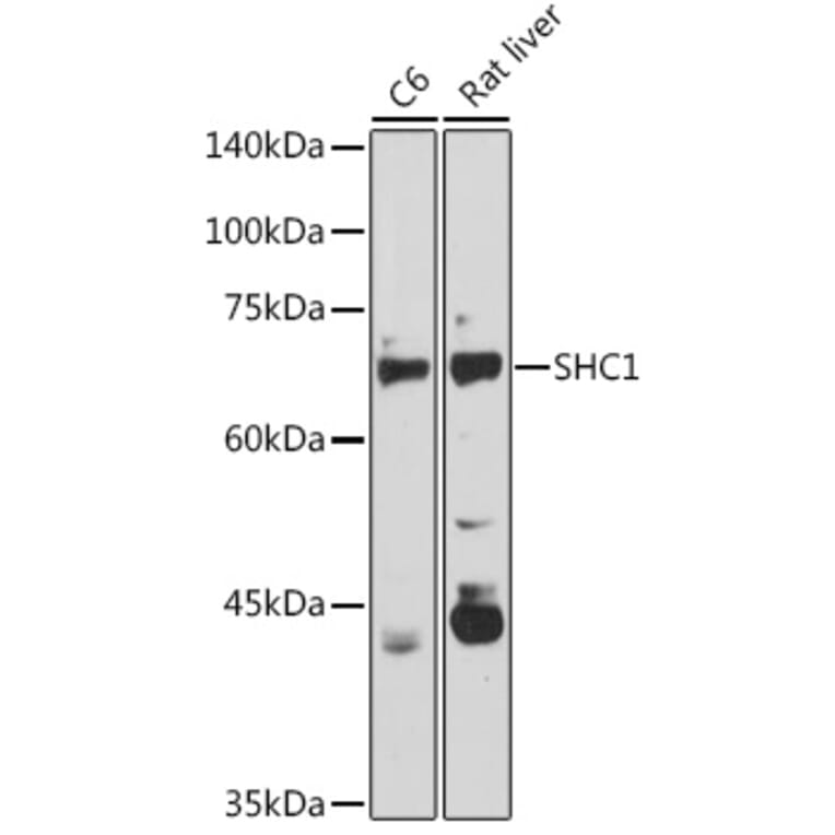
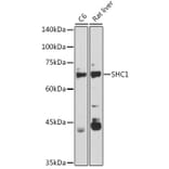
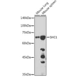
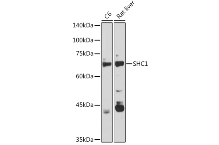
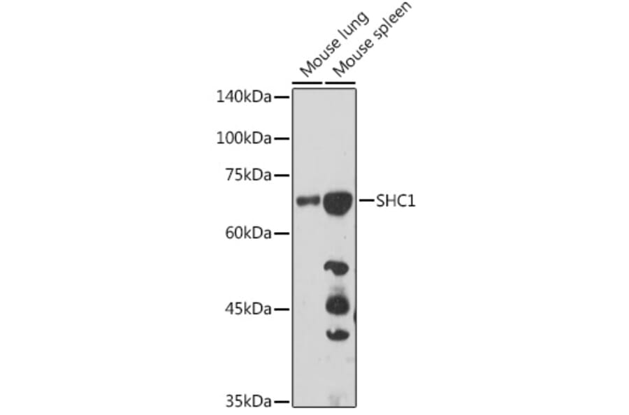
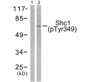
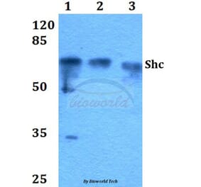
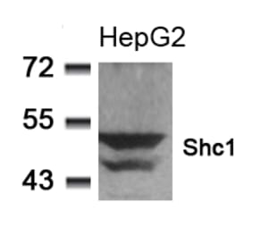
Western blot analysis of extracts of various cell lines, using Anti-SHC Antibody (A16912) at 1:1,000 dilution. The secondary antibody was Goat Anti-Rabbit IgG H&L Antibody (HRP) at 1:10,000 dilution. Lysates/proteins were present at 25µg per lane. The blocking buffer used was 3% non-fat dry milk in TBST. Detection was with a ECL Basic Kit. Exposure time: 90s.
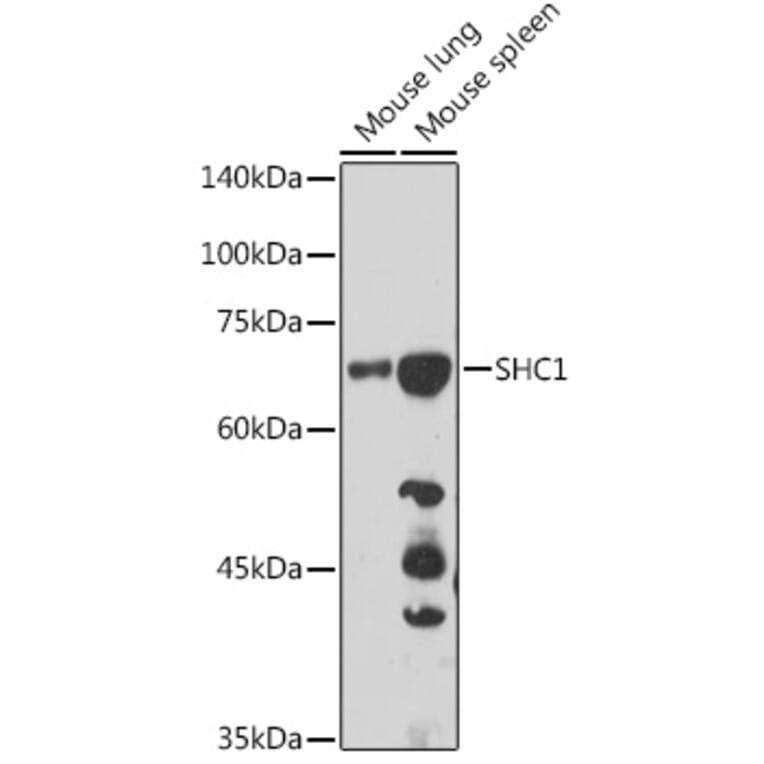
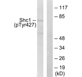
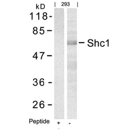
Western blot analysis of extracts of various cell lines, using Anti-SHC Antibody (A16912) at 1:1,000 dilution. The secondary antibody was Goat Anti-Rabbit IgG H&L Antibody (HRP) at 1:10,000 dilution. Lysates/proteins were present at 25µg per lane. The blocking buffer used was 3% non-fat dry milk in TBST. Detection was with a ECL Basic Kit. Exposure time: 180s.
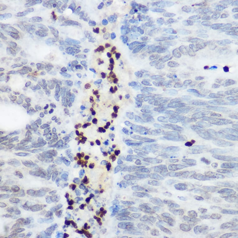
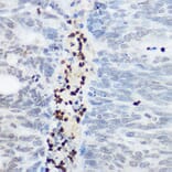
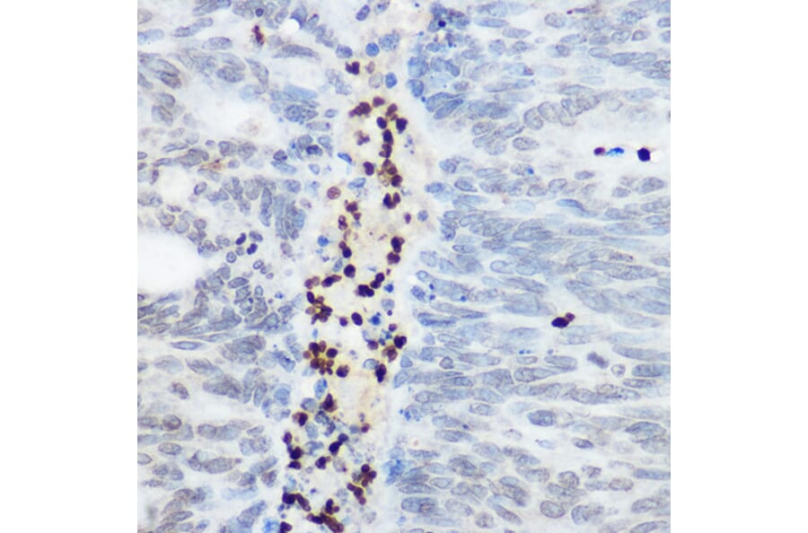
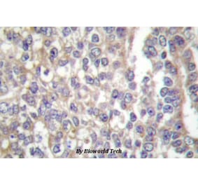
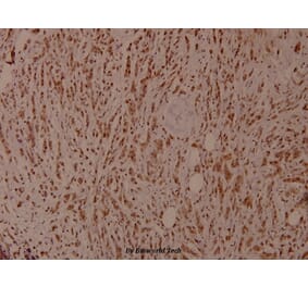
Immunohistochemistry analysis of paraffin-embedded human colon carcinoma tissue using Anti-SHC Antibody (A16912) at a dilution of 1:100 (40x lens). Perform high pressure antigen retrieval with 10 mM citrate buffer pH 6.0 before commencing with IHC staining protocol.
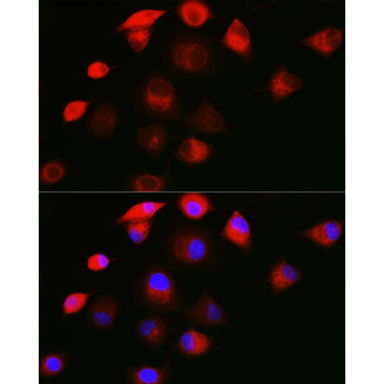
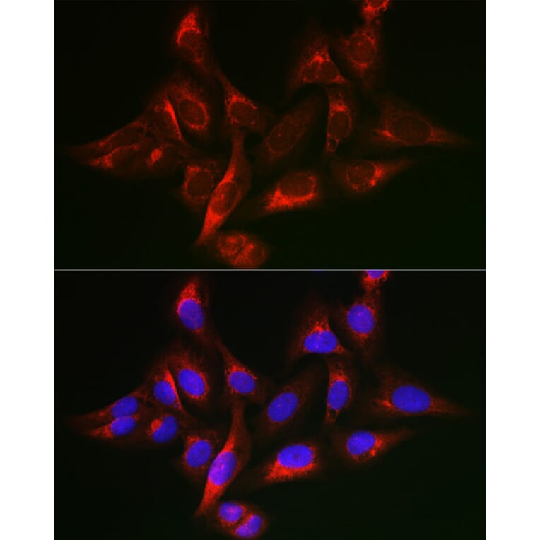
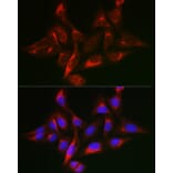
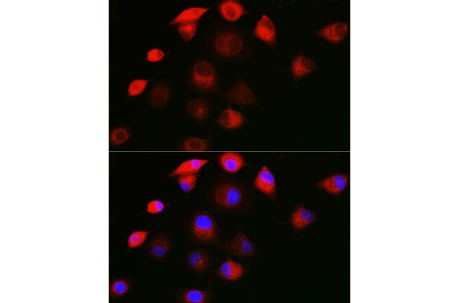
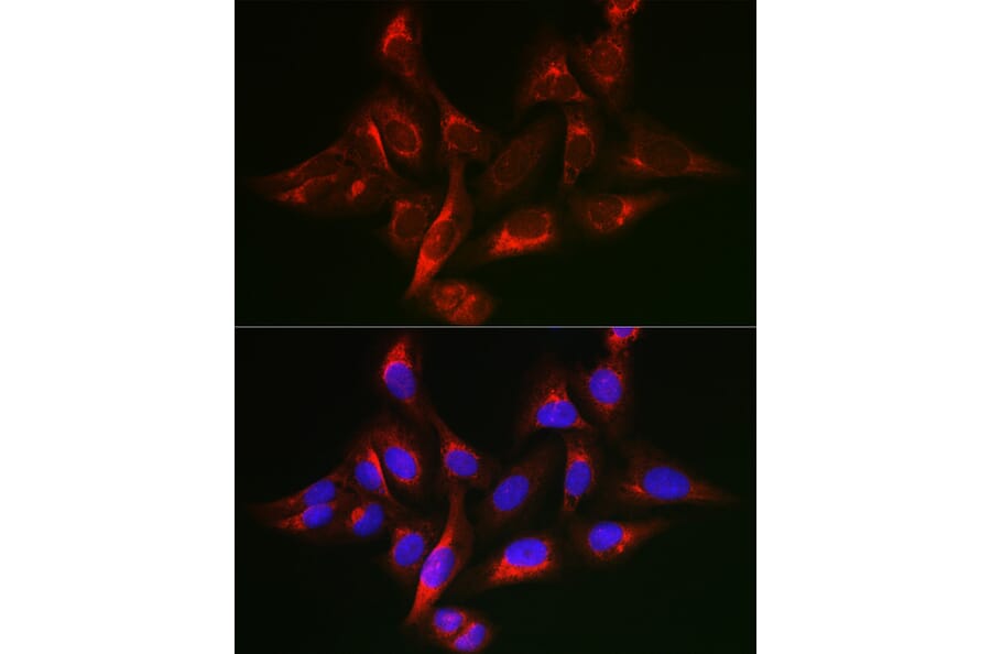
Immunofluorescence analysis of A549 cells using Anti-SHC Antibody (A16912) at a dilution of 1:50 (40x lens). DAPI was used to stain the cell nuclei (blue).
Immunofluorescence analysis of U-2 OS cells using Anti-SHC Antibody (A16912) at a dilution of 1:50 (40x lens). DAPI was used to stain the cell nuclei (blue).